
Search Count: 11
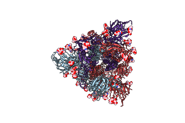 |
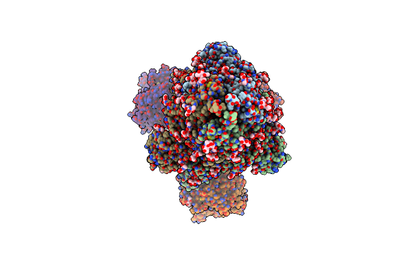
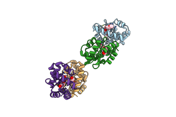
Cryo-Em Structure Of Sars-Cov-2 S-Uk Variant (B.1.1.7), One Rbd-Up Conformation 1
Organism: Severe acute respiratory syndrome coronavirus 2
Method: ELECTRON MICROSCOPY Release Date: 2021-09-01 Classification: VIRAL PROTEIN Ligands: NAG |
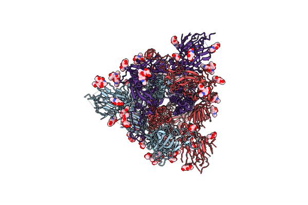 |
Cryo-Em Structure Of Sars-Cov-2 S-Uk Variant (B.1.1.7), One Rbd-Up Conformation 2
Organism: Severe acute respiratory syndrome coronavirus 2
Method: ELECTRON MICROSCOPY Release Date: 2021-09-01 Classification: VIRAL PROTEIN Ligands: NAG |
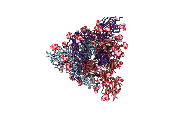 |
Cryo-Em Structure Of Sars-Cov-2 S-Uk Variant (B.1.1.7), One Rbd-Up Conformation 3
Organism: Severe acute respiratory syndrome coronavirus 2
Method: ELECTRON MICROSCOPY Release Date: 2021-09-01 Classification: VIRAL PROTEIN Ligands: NAG |
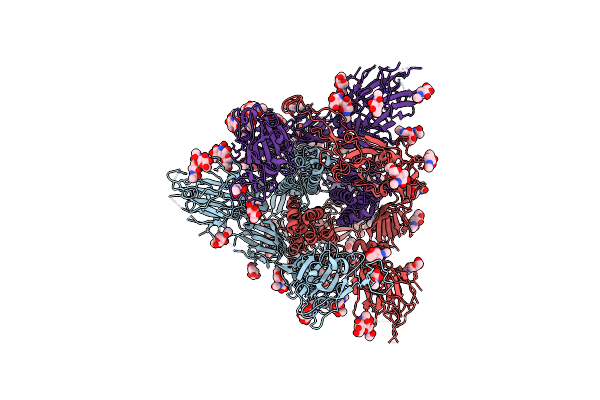 |
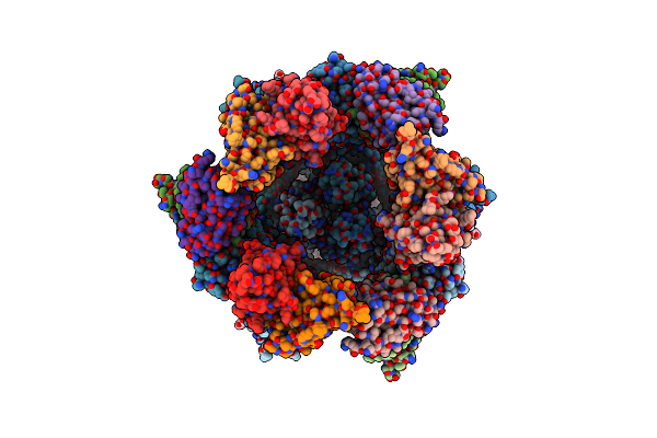
Cryo-Em Structure Of Sars-Cov-2 S-Uk Variant (B.1.1.7), Two Rbd-Up Conformation
Organism: Severe acute respiratory syndrome coronavirus 2
Method: ELECTRON MICROSCOPY Release Date: 2021-09-01 Classification: VIRAL PROTEIN Ligands: NAG |
 |
Cryo-Em Structure Of Sars-Cov-2 S-Uk Variant (B.1.1.7) In Complex With Angiotensin-Converting Enzyme 2 (Ace2) Ectodomain
Organism: Severe acute respiratory syndrome coronavirus 2, Homo sapiens
Method: ELECTRON MICROSCOPY Release Date: 2021-09-01 Classification: VIRAL PROTEIN Ligands: NAG |
 |
Cryo-Em Structure Of Sars-Cov-2 S-D614G Variant In Complex With Neutralizing Antibodies, Rbd-Chab15 And Rbd-Chab45
Organism: Severe acute respiratory syndrome coronavirus 2, Homo sapiens
Method: ELECTRON MICROSCOPY Release Date: 2021-09-01 Classification: VIRAL PROTEIN Ligands: NAG |
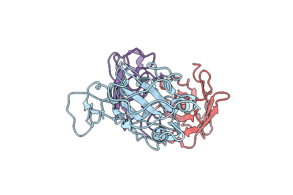 |
Cryo-Em Structure Of Sars-Cov-2 Spike In Complex With A Neutralizing Antibody Chab-25 (Focused Refinement Of S-Rbd And Chab-25 Region)
Organism: Severe acute respiratory syndrome coronavirus 2, Homo sapiens
Method: ELECTRON MICROSCOPY Release Date: 2021-08-04 Classification: VIRAL PROTEIN |
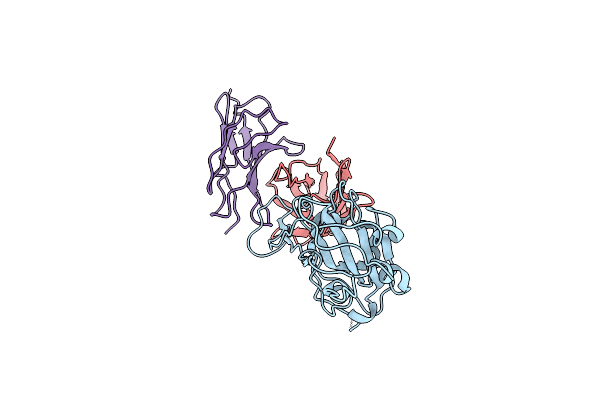 |
Cryo-Em Structure Of Sars-Cov-2 Spike In Complex With A Neutralizing Antibody Chab-45 (Focused Refinement Of S-Rbd And Chab-45 Region)
Organism: Severe acute respiratory syndrome coronavirus 2, Homo sapiens
Method: ELECTRON MICROSCOPY Release Date: 2021-08-04 Classification: VIRAL PROTEIN |
 |
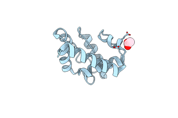
Structure Of Anopheles Gambiae Odorant Binding Protein 20 Bound To Polyethylene Glycol
Organism: Anopheles gambiae
Method: X-RAY DIFFRACTION Resolution:1.80 Å Release Date: 2012-10-17 Classification: ODORANT-BINDING PROTEIN Ligands: PG4 |
 |
Crystal Structure Of Anopholes Gambiae Odorant Binding Protein 20 In Open State
Organism: Anopheles gambiae
Method: X-RAY DIFFRACTION Resolution:2.00 Å Release Date: 2012-10-17 Classification: ODORANT-BINDING PROTEIN Ligands: ACY |
 |
Organism: Anopheles gambiae
Method: X-RAY DIFFRACTION Resolution:1.80 Å Release Date: 2012-10-17 Classification: ODORANT-BINDING PROTEIN Ligands: 2PE, PG6 |