
Search Count: 23
 |
Organism: Mus musculus
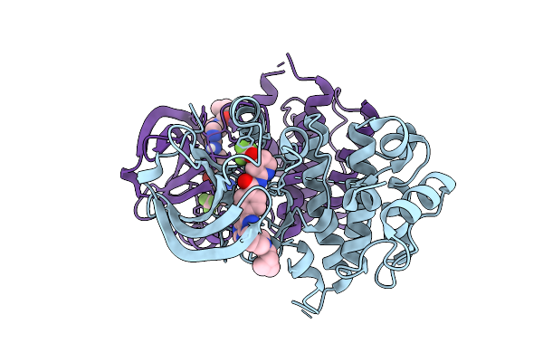
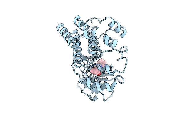
Method: X-RAY DIFFRACTION Resolution:2.03 Å Release Date: 2025-08-13 Classification: TRANSFERASE/TRANSFERASE INHIBITOR Ligands: A1BIZ |
 |
Organism: Mus musculus
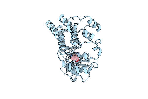
Method: X-RAY DIFFRACTION Resolution:2.10 Å Release Date: 2025-08-13 Classification: TRANSFERASE/TRANSFERASE INHIBITOR Ligands: A1BI0 |
 |
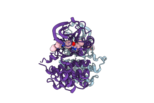
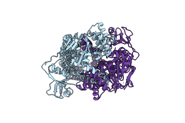
Bruton'S Tyrosine Kinase With Mutations In The Activation Loop In Complex With Compound Pti52
Organism: Mus musculus
Method: X-RAY DIFFRACTION Resolution:2.26 Å Release Date: 2025-08-13 Classification: TRANSFERASE/TRANSFERASE INHIBITOR Ligands: A1BI0, DMS |
 |
Bruton'S Tyrosine Kinase With Mutations In The Activation Loop In Complex With Compound Pti42
Organism: Mus musculus
Method: X-RAY DIFFRACTION Resolution:2.86 Å Release Date: 2025-08-13 Classification: TRANSFERASE/TRANSFERASE INHIBITOR Ligands: A1BI1 |
 |
Bruton'S Tyrosine Kinase With Mutations In The Activation Loop In Complex With Compound A110162
Organism: Mus musculus
Method: X-RAY DIFFRACTION Resolution:3.38 Å Release Date: 2025-08-13 Classification: TRANSFERASE/TRANSFERASE INHIBITOR Ligands: A1BKW |
 |
Bruton'S Tyrosine Kinase With Mutations In The Activation Loop In Complex With Compound P301390
Organism: Mus musculus
Method: X-RAY DIFFRACTION Resolution:3.05 Å Release Date: 2025-08-13 Classification: TRANSFERASE/TRANSFERASE INHIBITOR Ligands: A1BJE |
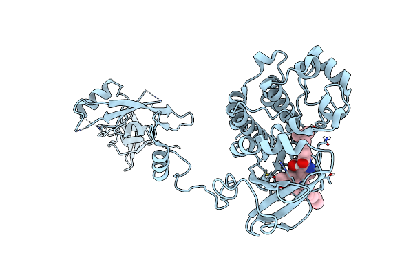 |
Crystal Structure Of The Full-Length Bruton'S Tyrosine Kinase (Ph-Th Domain Not Visible)
Organism: Mus musculus
Method: X-RAY DIFFRACTION Resolution:3.40 Å Release Date: 2023-08-16 Classification: TRANSFERASE Ligands: 9AJ |
 |
Organism: Mus musculus
Method: X-RAY DIFFRACTION Resolution:2.10 Å Release Date: 2023-08-16 Classification: TRANSFERASE Ligands: 9AJ, ZN, GOL |
 |
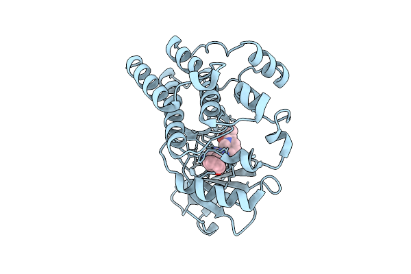
Crystal Structure Of The Kinase Domain Of Bruton'S Tyrosine Kinase Bound To Dasatinib
Organism: Mus musculus
Method: X-RAY DIFFRACTION Resolution:2.60 Å Release Date: 2023-08-16 Classification: TRANSFERASE Ligands: 1N1 |
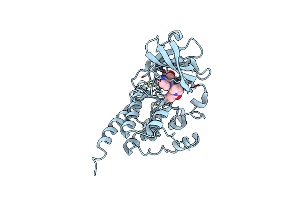 |
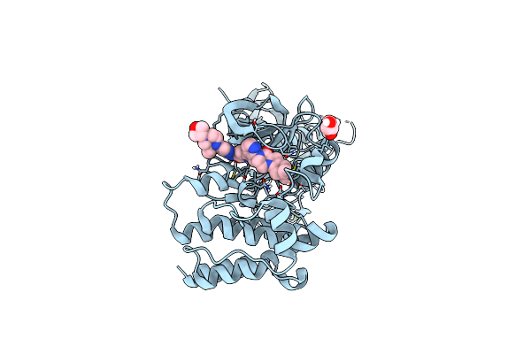
Structure Of Btk Kinase Domain With The Second-Generation Inhibitor Acalabrutinib
Organism: Mus musculus
Method: X-RAY DIFFRACTION Resolution:1.70 Å Release Date: 2023-07-05 Classification: TRANSFERASE/TRANSFERASE INHIBITOR Ligands: XQQ, BR |
 |
Structure Of Btk Kinase Domain With The Second-Generation Inhibitor Tirabrutinib
Organism: Mus musculus
Method: X-RAY DIFFRACTION Resolution:2.60 Å Release Date: 2023-07-05 Classification: TRANSFERASE/TRANSFERASE INHIBITOR Ligands: 7GB |
 |
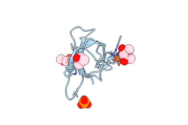
Crystal Structure Of Trimethoprim-Resistant Type Ii Dihydrofolate Reductase In Complex With A Bisbenzimidazole Inhibitor
Organism: Escherichia coli
Method: X-RAY DIFFRACTION Resolution:1.75 Å Release Date: 2019-05-29 Classification: OXIDOREDUCTASE Ligands: D49, MRD, PO4 |
 |
Crystal Structure Of Trimethoprim-Resistant Type Ii Dihydrofolate Reductase In Complex With A Bisbenzimidazole Inhibitor
Organism: Escherichia coli
Method: X-RAY DIFFRACTION Resolution:1.40 Å Release Date: 2019-05-29 Classification: OXIDOREDUCTASE Ligands: LBA, MRD, PO4 |
 |
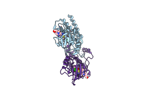
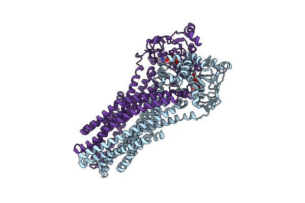
Organism: Ruminiclostridium thermocellum atcc 27405
Method: X-RAY DIFFRACTION Resolution:3.61 Å Release Date: 2015-07-22 Classification: TRANSPORT PROTEIN/HYDROLASE |
 |
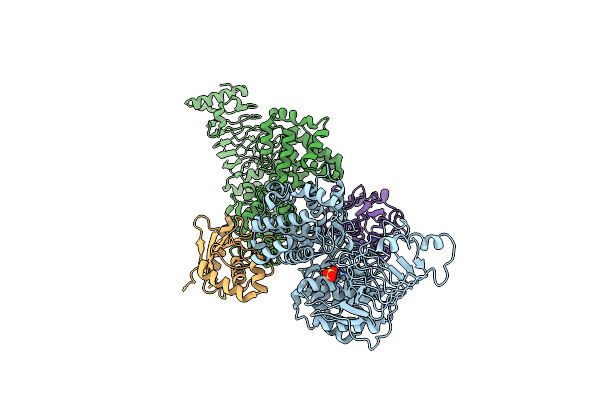
Crystal Structure Of The Peptidase-Containing Abc Transporter Pcat1 E648Q Mutant Complexed With Atpgs In An Occluded Conformation
Organism: Ruminiclostridium thermocellum atcc 27405
Method: X-RAY DIFFRACTION Resolution:5.52 Å Release Date: 2015-07-22 Classification: TRANSPORT PROTEIN/HYDROLASE Ligands: AGS |
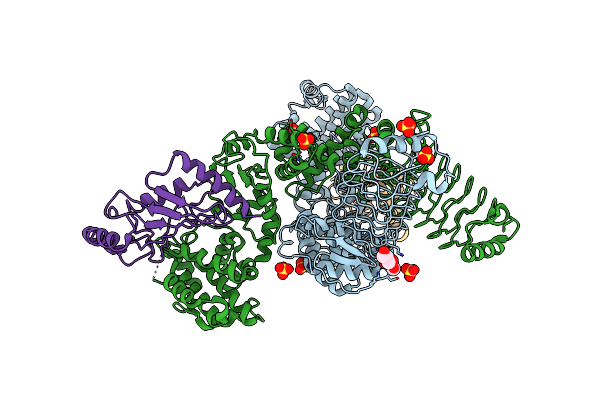 |
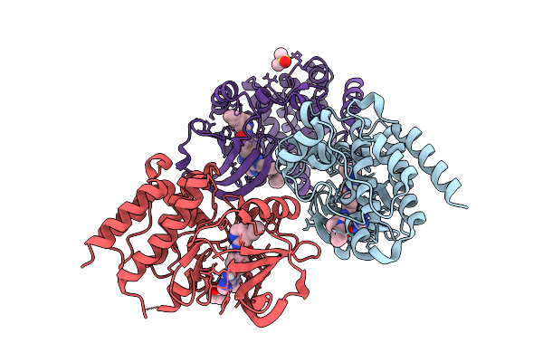
Crystal Structure Of E. Coli O157:H7 E3 Ubiquitin Ligase, Nlel, With A Human E2, Ubch7
Organism: Escherichia coli, Homo sapiens
Method: X-RAY DIFFRACTION Resolution:3.30 Å Release Date: 2012-01-25 Classification: LIGASE/SIGNALING PROTEIN Ligands: SO4, GOL |
 |
Crystal Structure Of The Salmonella E3 Ubiquitin Ligase Sopa In Complex With The Human E2 Ubch7
Organism: Salmonella enterica subsp. enterica serovar typhimurium, Homo sapiens
Method: X-RAY DIFFRACTION Resolution:3.27 Å Release Date: 2012-01-25 Classification: LIGASE/SIGNALING PROTEIN Ligands: SO4 |
 |
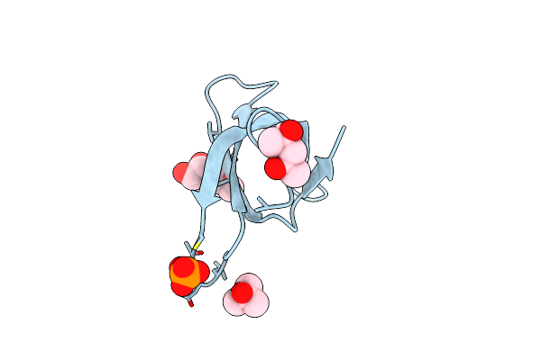
Novel Crystallization Conditions For Tandem Variant R67 Dhfr Yields Wild-Type Crystal Structure
Organism: Escherichia coli
Method: X-RAY DIFFRACTION Resolution:1.40 Å Release Date: 2011-11-02 Classification: OXIDOREDUCTASE Ligands: MRD |
 |
Organism: Escherichia coli
Method: X-RAY DIFFRACTION Resolution:2.50 Å Release Date: 2010-10-27 Classification: LIGASE Ligands: SO4, GOL, MES |
 |
Organism: Escherichia coli
Method: X-RAY DIFFRACTION Resolution:2.10 Å Release Date: 2010-10-27 Classification: LIGASE Ligands: GOL, MES, SO4, DTT |