
Search Count: 34
 |
Organism: Escherichia coli (strain k12), Bacteroides fragilis
Method: X-RAY DIFFRACTION Resolution:2.72 Å Release Date: 2025-10-01 Classification: TOXIN |
 |
Organism: Bacteroides fragilis
Method: X-RAY DIFFRACTION Resolution:2.00 Å Release Date: 2025-10-01 Classification: ANTITOXIN Ligands: IPA |
 |
Organism: Bacteroides fragilis nctc 9343
Method: X-RAY DIFFRACTION Resolution:1.90 Å Release Date: 2025-10-01 Classification: STRUCTURAL PROTEIN |
 |
Structure Of A De Novo Designed Interleukin-21 Mimetic Complex With Il-21R And Il-2Rg
Organism: Synthetic construct, Homo sapiens
Method: X-RAY DIFFRACTION Resolution:2.28 Å Release Date: 2025-09-24 Classification: DE NOVO PROTEIN/IMMUNE SYSTEM Ligands: NAG, EDO |
 |
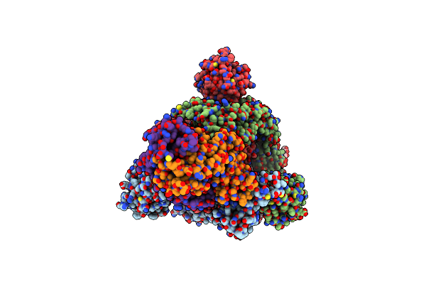
Structure Of Cargo Complex (Btpea-Btaeb-Btapc) Bound To The Vgrg Spike From The Type Vi Secretion System
Organism: Bacteroides fragilis
Method: ELECTRON MICROSCOPY Release Date: 2025-09-10 Classification: ANTIMICROBIAL PROTEIN Ligands: ZN |
 |
Organism: Bacteroides fragilis
Method: X-RAY DIFFRACTION Resolution:2.18 Å Release Date: 2025-09-03 Classification: ANTIMICROBIAL PROTEIN Ligands: PEG |
 |
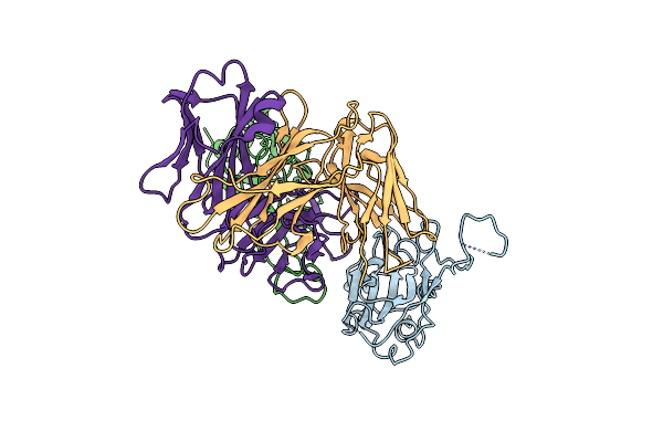
Crystal Structure Of The Type Vi Secretion System Effector-Immunity Complex Btpea Ctd-Btpia From Bacteroides Fragilis.
Organism: Bacteroides fragilis
Method: X-RAY DIFFRACTION Resolution:2.11 Å Release Date: 2025-09-03 Classification: ANTIMICROBIAL PROTEIN Ligands: GOL |
 |
Organism: Escherichia coli k-12, Bacteroides fragilis nctc 9343
Method: X-RAY DIFFRACTION Resolution:2.40 Å Release Date: 2025-02-12 Classification: ANTITOXIN Ligands: GOL |
 |
Organism: Bacteroides thetaiotaomicron
Method: X-RAY DIFFRACTION Resolution:3.00 Å Release Date: 2025-02-12 Classification: CHAPERONE Ligands: PEG |
 |
Organism: Bacteroides fragilis
Method: ELECTRON MICROSCOPY Release Date: 2024-09-25 Classification: MEMBRANE PROTEIN |
 |
Organism: Bacteroides fragilis
Method: ELECTRON MICROSCOPY Release Date: 2024-09-25 Classification: MEMBRANE PROTEIN |
 |
Crystal Structure Of Human Ubiquitin-Like Protein From Bacteroides Fragilis
Organism: Bacteroides fragilis
Method: X-RAY DIFFRACTION Resolution:1.29 Å Release Date: 2023-11-29 Classification: CELL CYCLE Ligands: EDO |
 |
Crystal Structure Of Human Ubiquitin-Like Protein From Bacteroides Fragilis C Terminal Cysteine Mutant
Organism: Bacteroides fragilis
Method: X-RAY DIFFRACTION Resolution:1.34 Å Release Date: 2023-11-29 Classification: CELL CYCLE |
 |
Organism: Bacteroides fragilis
Method: X-RAY DIFFRACTION Resolution:2.26 Å Release Date: 2023-11-29 Classification: ISOMERASE Ligands: GOL, SO4, MG |
 |
Organism: Bacteroides fragilis
Method: X-RAY DIFFRACTION Resolution:3.79 Å Release Date: 2023-11-29 Classification: ISOMERASE |
 |
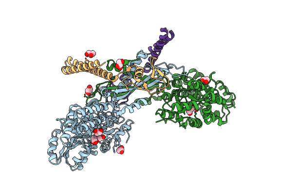
Sars-Cov-2 Spike Glycoprotein Trimer Complexed With Fab Fragment Of Anti-Rbd Antibody E7
Organism: Homo sapiens
Method: ELECTRON MICROSCOPY Release Date: 2023-08-02 Classification: VIRAL PROTEIN Ligands: NAG |
 |
Sars-Cov-2 Spike Glycoprotein Trimer Complexed With Fab Fragment Of Anti-Rbd Antibody E7 (Focused Refinement On Fab-Rbd Interface)
Organism: Homo sapiens
Method: ELECTRON MICROSCOPY Release Date: 2023-08-02 Classification: VIRAL PROTEIN |
 |
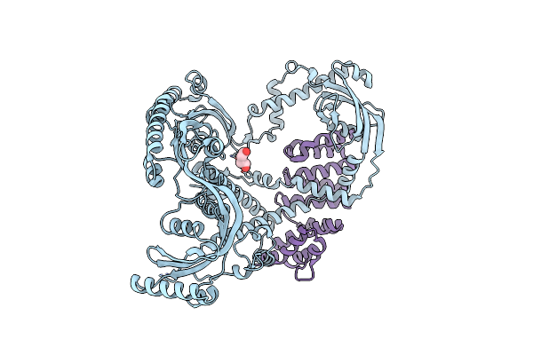
The Crystal Structure Of The Tir Domain-Containing Protein From Acinetobacter Baumannii (Abtir)
Organism: Acinetobacter baumannii
Method: X-RAY DIFFRACTION Resolution:2.16 Å Release Date: 2022-09-07 Classification: HYDROLASE Ligands: SO4, P6G |
 |
Organism: Bacteroides thetaiotaomicron
Method: X-RAY DIFFRACTION Resolution:1.42 Å Release Date: 2022-09-07 Classification: HYDROLASE |
 |
Organism: Bacillus subtilis
Method: X-RAY DIFFRACTION Resolution:1.57 Å Release Date: 2022-09-07 Classification: HYDROLASE Ligands: OJC, GOL, SO4 |