
Search Count: 16
 |
Organism: Homo sapiens
Method: ELECTRON MICROSCOPY Release Date: 2024-10-02 Classification: HYDROLASE |
 |
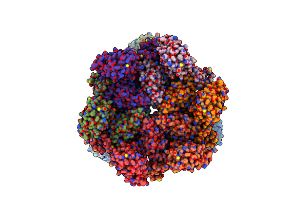
Organism: Bacillus subtilis
Method: ELECTRON MICROSCOPY Release Date: 2024-09-11 Classification: IMMUNE SYSTEM |
 |
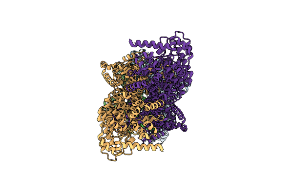
Organism: Bacillus subtilis
Method: ELECTRON MICROSCOPY Release Date: 2024-09-11 Classification: IMMUNE SYSTEM |
 |
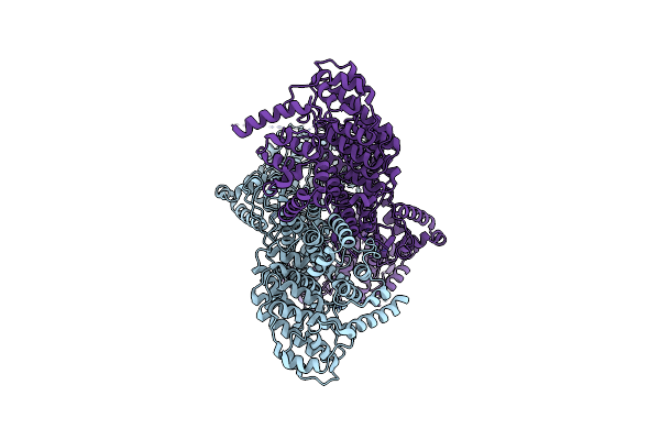
Organism: Bacillus subtilis
Method: ELECTRON MICROSCOPY Release Date: 2024-09-11 Classification: IMMUNOSUPPRESSANT |
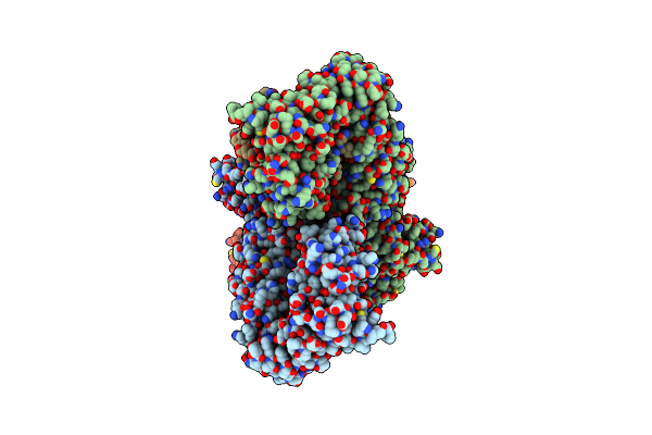 |
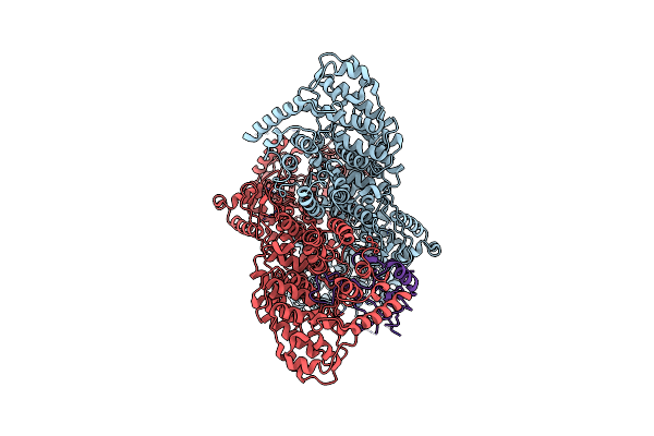
The Cryo-Em Structure Of Anti-Phage Defense Associated Dsr2 Tetramer Bound With Two Dsad1 Inhibitors (Same Side)
Organism: Bacillus subtilis
Method: ELECTRON MICROSCOPY Release Date: 2024-09-11 Classification: IMMUNOSUPPRESSANT |
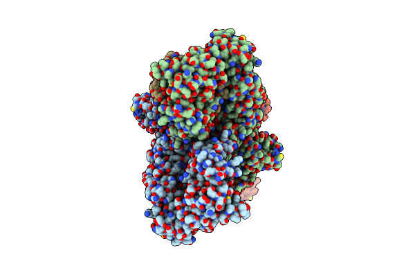 |
The Cryo-Em Structure Of Anti-Phage Defense Associated Dsr2 Tetramer Bound With Two Dsad1 Inhibitors (Opposite Side)
Organism: Bacillus subtilis
Method: ELECTRON MICROSCOPY Release Date: 2024-09-11 Classification: IMMUNOSUPPRESSANT |
 |
Organism: Bacillus subtilis
Method: ELECTRON MICROSCOPY Release Date: 2024-09-11 Classification: IMMUNE SYSTEM Ligands: NAD |
 |
Organism: Arabidopsis thaliana
Method: X-RAY DIFFRACTION Resolution:3.26 Å Release Date: 2024-02-07 Classification: PLANT PROTEIN Ligands: FE |
 |
Organism: Homo sapiens
Method: X-RAY DIFFRACTION Resolution:1.88 Å Release Date: 2021-02-24 Classification: TRANSFERASE Ligands: SO4 |
 |
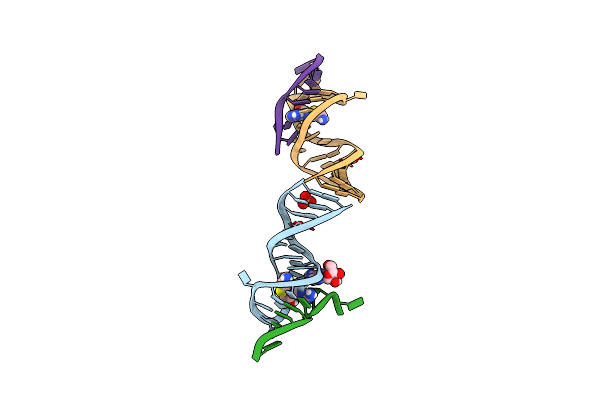
Organism: Roseobacter sp.
Method: X-RAY DIFFRACTION Release Date: 2020-07-22 Classification: RNA Ligands: SAH, MG, NA, SO4 |
 |
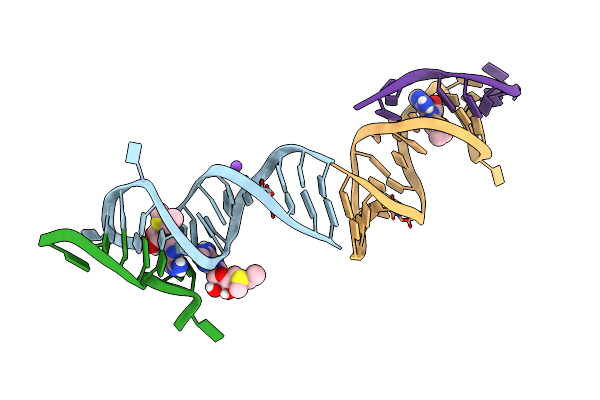
Organism: Roseobacter sp.
Method: X-RAY DIFFRACTION Resolution:2.12 Å Release Date: 2020-07-22 Classification: RNA Ligands: SAM |
 |
Organism: Roseobacter sp.
Method: X-RAY DIFFRACTION Resolution:2.50 Å Release Date: 2020-07-22 Classification: RNA Ligands: CBV, AMP |
 |
Organism: Roseobacter sp.
Method: X-RAY DIFFRACTION Resolution:2.04 Å Release Date: 2020-07-22 Classification: RNA Ligands: ADN, NA, CBV, MG |
 |
Organism: Roseobacter sp.
Method: X-RAY DIFFRACTION Resolution:2.03 Å Release Date: 2020-07-22 Classification: RNA Ligands: MTA, SO4, NA |
 |
Organism: Roseobacter sp.
Method: X-RAY DIFFRACTION Resolution:2.17 Å Release Date: 2020-07-22 Classification: RNA Ligands: AMP, DSH, SO4 |
 |
Organism: Roseobacter sp.
Method: X-RAY DIFFRACTION Resolution:2.20 Å Release Date: 2020-07-22 Classification: RNA Ligands: SAM, NA |