
Search Count: 28
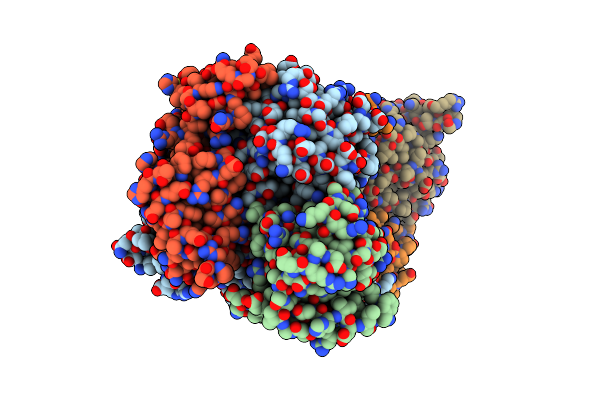 |
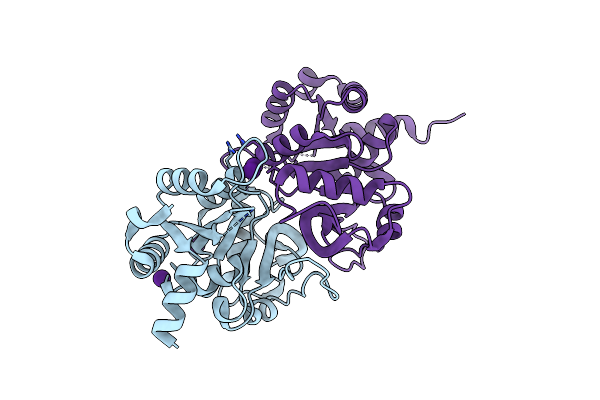
Organism: Klebsiella pneumoniae
Method: X-RAY DIFFRACTION Resolution:2.50 Å Release Date: 2003-06-10 Classification: LYASE Ligands: K, B12 |
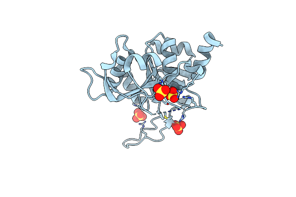 |
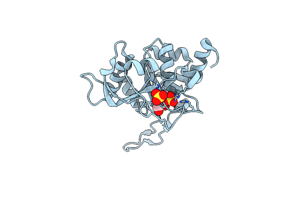
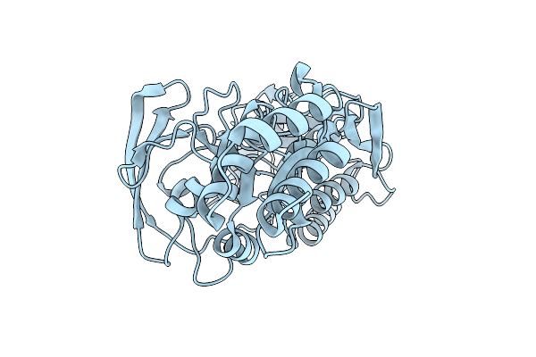
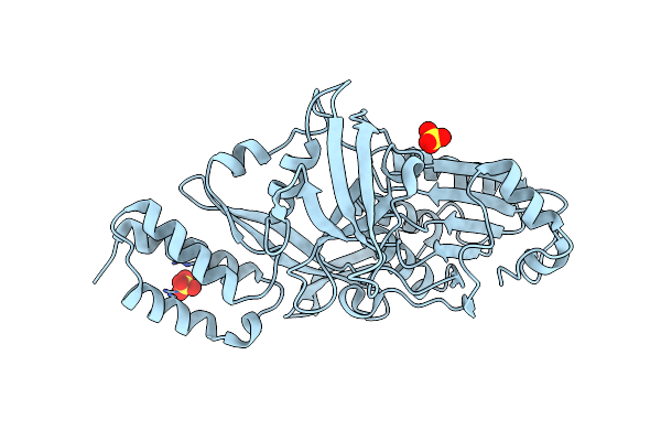
Crystal Structure Of 3,4-Dihydroxy-2-Butanone 4-Phosphate Synthase (Cation Free Form)
Organism: Magnaporthe grisea
Method: X-RAY DIFFRACTION Resolution:1.50 Å Release Date: 2002-03-06 Classification: ISOMERASE Ligands: SO4 |
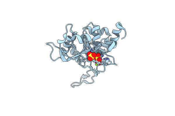 |
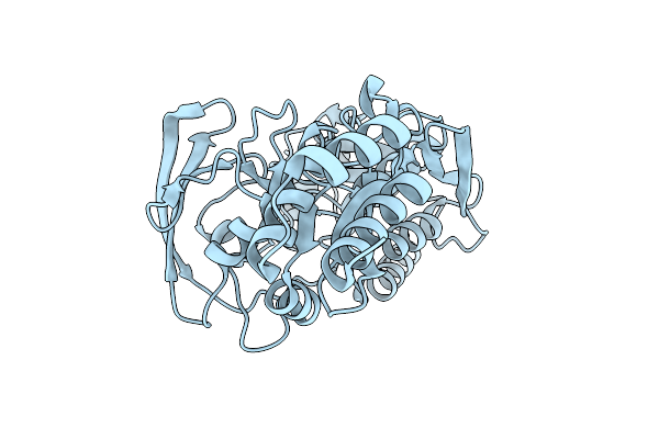
Crystal Structure Of 3,4-Dihydroxy-2-Butanone 4-Phosphate Synthase In Complex With Two Magnesium Ions
Organism: Magnaporthe grisea
Method: X-RAY DIFFRACTION Resolution:0.98 Å Release Date: 2002-03-06 Classification: ISOMERASE Ligands: SO4, MG |
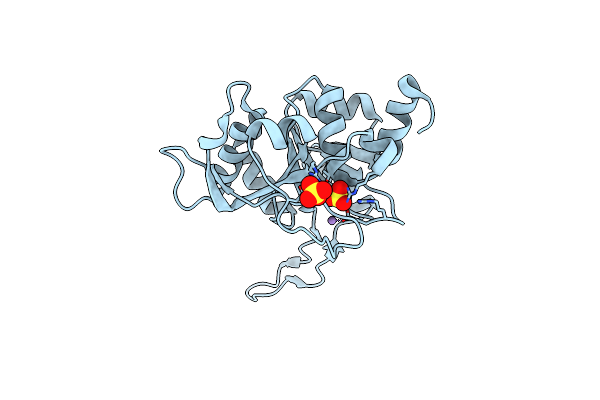 |
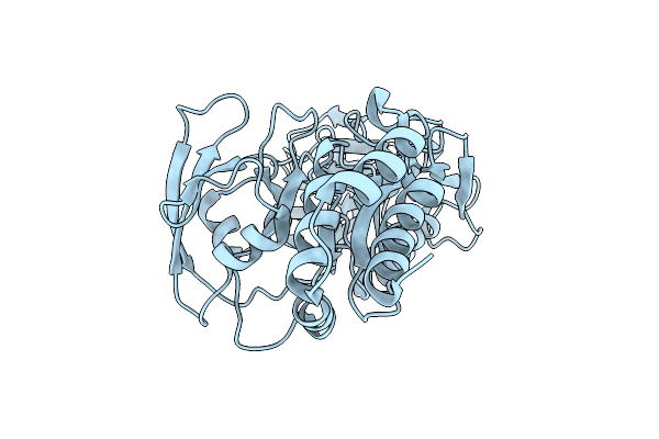
Crystal Structure Of 3,4-Dihydroxy-2-Butanone 4-Phosphate Synthase In Complex With Two Manganese Ions
Organism: Magnaporthe grisea
Method: X-RAY DIFFRACTION Resolution:1.60 Å Release Date: 2002-03-06 Classification: ISOMERASE Ligands: SO4, MN |
 |
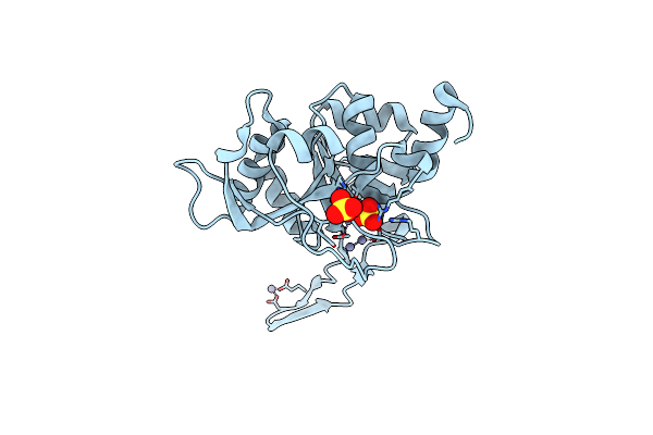
Crystal Structure Of 3,4-Dihydroxy-2-Butanone 4-Phosphate Synthase In Complex With One Manganese, And A Glycerol
Organism: Magnaporthe grisea
Method: X-RAY DIFFRACTION Resolution:1.10 Å Release Date: 2002-03-06 Classification: ISOMERASE Ligands: SO4, MN, GOL |
 |
Crystal Structure Of 3,4-Dihydroxy-2-Butanone 4-Phosphate Synthase In Complex With Zinc Ions
Organism: Magnaporthe grisea
Method: X-RAY DIFFRACTION Resolution:1.00 Å Release Date: 2002-03-06 Classification: ISOMERASE Ligands: SO4, ZN |
 |
Organism: Escherichia coli
Method: X-RAY DIFFRACTION Resolution:2.00 Å Release Date: 2001-09-19 Classification: TRANSFERASE |
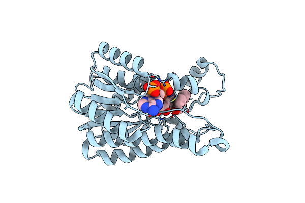 |
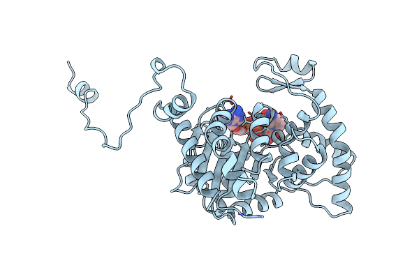
Crystal Structure Of 1,3,6,8-Tetrahydroxynaphthalene Reductase In Complex With Nadph And Pyroquilon
Organism: Magnaporthe grisea
Method: X-RAY DIFFRACTION Resolution:1.50 Å Release Date: 2001-09-19 Classification: OXIDOREDUCTASE Ligands: NDP, PYQ |
 |
Organism: Escherichia coli
Method: X-RAY DIFFRACTION Resolution:1.40 Å Release Date: 2001-04-30 Classification: ISOMERASE Ligands: CS |
 |
Crystal Structure Of 3,4-Dihydroxy-2-Butanone 4-Phosphate Synthase Gold Derivative
Organism: Escherichia coli
Method: X-RAY DIFFRACTION Resolution:1.55 Å Release Date: 2001-04-30 Classification: ISOMERASE Ligands: AU |
 |
Organism: Scenedesmus obliquus
Method: X-RAY DIFFRACTION Resolution:1.80 Å Release Date: 2001-01-18 Classification: HYDROLASE |
 |
Organism: Scenedesmus obliquus
Method: X-RAY DIFFRACTION Resolution:2.00 Å Release Date: 2001-01-18 Classification: HYDROLASE |
 |
Organism: Scenedesmus obliquus
Method: X-RAY DIFFRACTION Resolution:1.90 Å Release Date: 2001-01-18 Classification: HYDROLASE |
 |
Organism: Scenedesmus obliquus
Method: X-RAY DIFFRACTION Resolution:2.10 Å Release Date: 2001-01-18 Classification: HYDROLASE Ligands: SO4 |
 |
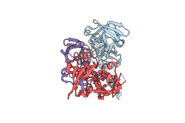
Organism: Homo sapiens
Method: X-RAY DIFFRACTION Resolution:2.20 Å Release Date: 1998-02-25 Classification: TRANSFERASE Ligands: NAD |
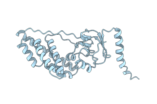 |
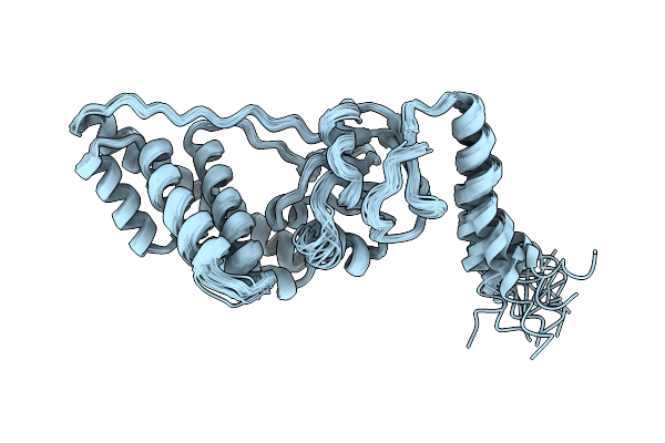
Amino Terminal Domain Of Enzyme I From Escherichia Coli Nmr, Restrained Regularized Mean Structure
Organism: Escherichia coli
Method: SOLUTION NMR Release Date: 1998-01-07 Classification: PHOSPHOTRANSFERASE |
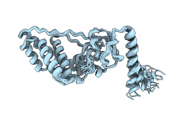 |
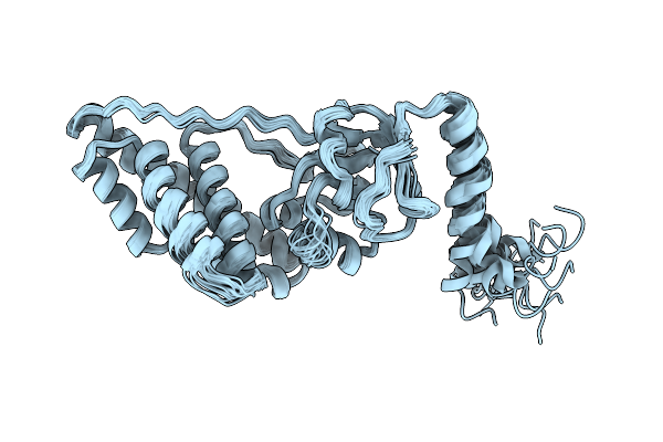
Amino Terminal Domain Of Enzyme I From Escherichia Coli, Nmr, 17 Structures
Organism: Escherichia coli
Method: SOLUTION NMR Release Date: 1998-01-07 Classification: PHOSPHOTRANSFERASE |
 |
Amino Terminal Domain Of Enzyme I From Escherichia Coli, Nmr, 17 Structures
Organism: Escherichia coli
Method: SOLUTION NMR Release Date: 1998-01-07 Classification: PHOSPHOTRANSFERASE |
 |
Organism: Escherichia coli
Method: SOLUTION NMR Release Date: 1998-01-07 Classification: PHOSPHOTRANSFERASE |
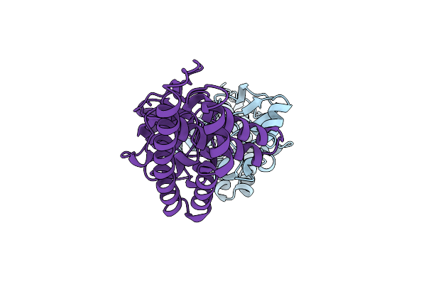 |
Organism: Escherichia coli
Method: X-RAY DIFFRACTION Resolution:2.50 Å Release Date: 1996-12-07 Classification: PHOSPHOTRANSFERASE |