
Search Count: 1,005
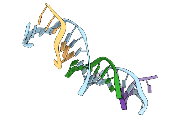 |
[T:Ag+/Hg2+:T--(Ph8-Ph9.5; 50S)] Metal-Mediated Dna Base Pair In Tensegrity Triangle Grown At Ph 8 And Soaked In Ph 9.5 Ag/Hg For 50S
Organism: Synthetic construct
Method: X-RAY DIFFRACTION Release Date: 2025-12-31 Classification: DNA Ligands: HG, AG |
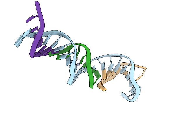 |
[T:Ag+/Hg2+:T--(Ph8-Ph9.5; 75S)] Metal-Mediated Dna Base Pair In Tensegrity Triangle Grown At Ph 8 And Soaked In Ph 9.5 For 75S
Organism: Synthetic construct
Method: X-RAY DIFFRACTION Release Date: 2025-12-31 Classification: DNA Ligands: HG, AG |
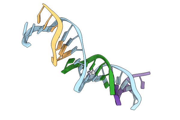 |
[T:Ag+/Hg2+:T--(Ph8-Ph9.5; 95S)] Metal-Mediated Dna Base Pair In Tensegrity Triangle Grown At Ph 8 And Soaked In Ph 9.5 Ag/Hg For 95S
Organism: Synthetic construct
Method: X-RAY DIFFRACTION Release Date: 2025-12-31 Classification: DNA Ligands: HG, AG |
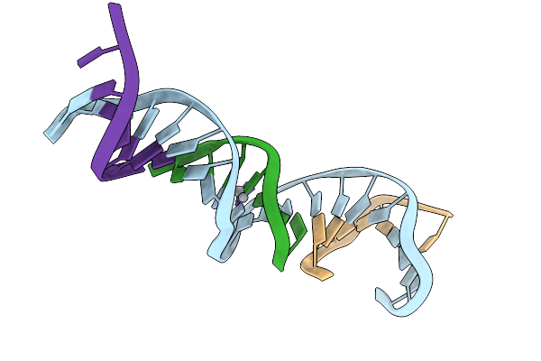 |
[T:Ag+/Hg2+:T--(Ph11-Ph9.5; 5S)] Metal-Mediated Dna Base Pair In Tensegrity Triangle Grown At Ph 8 And Soaked In Ph 9.5 Ag/Hg For 5S
Organism: Synthetic construct
Method: X-RAY DIFFRACTION Release Date: 2025-12-31 Classification: DNA Ligands: AG, HG |
 |
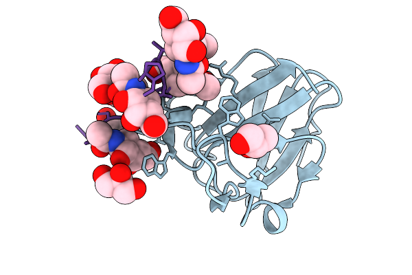
Organism: Escherichia coli, Synthetic construct
Method: X-RAY DIFFRACTION Release Date: 2025-12-24 Classification: SUGAR BINDING PROTEIN Ligands: A2G |
 |
Organism: Escherichia coli, Synthetic construct
Method: X-RAY DIFFRACTION Release Date: 2025-12-24 Classification: SUGAR BINDING PROTEIN Ligands: GOL, A2G |
 |
Organism: Escherichia coli, Synthetic construct
Method: X-RAY DIFFRACTION Release Date: 2025-12-24 Classification: SUGAR BINDING PROTEIN |
 |
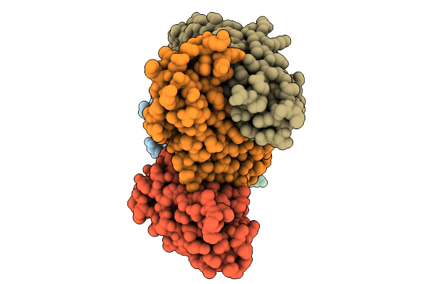
Organism: Monkeypox virus, Homo sapiens
Method: ELECTRON MICROSCOPY Release Date: 2025-12-24 Classification: VIRAL PROTEIN |
 |
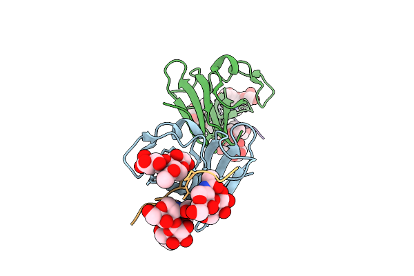
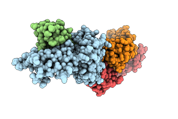
Globular Domain Of Monkeypox Virus Opg153 (A28) In Complex With Antibodies 08E11 And 12I12
Organism: Monkeypox virus, Homo sapiens
Method: ELECTRON MICROSCOPY Release Date: 2025-12-24 Classification: VIRAL PROTEIN |
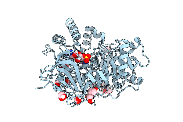 |
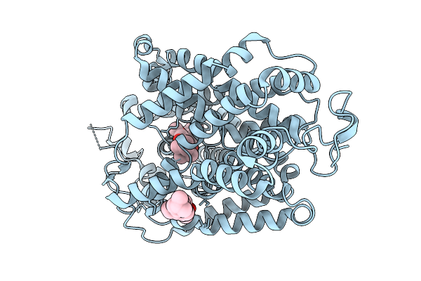
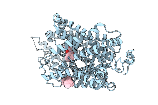
High-Resolution Crystal Structure Of Methyl-2,3-Diamino Propanoic Acid-Ams Inhibitor Bound Adenylation Domain (A3) From Sulfazecin Nonribosomal Peptide Synthetase Sulm
Organism: Paraburkholderia acidicola
Method: X-RAY DIFFRACTION Release Date: 2025-12-10 Classification: LIGASE Ligands: A1BVA, EDO, PEG |
 |
Organism: Xenopus tropicalis
Method: ELECTRON MICROSCOPY Release Date: 2025-11-19 Classification: MEMBRANE PROTEIN |
 |
Cryo-Em Structure Of Glycine Transporter 2 In Complex With Substrate Glycine
Organism: Xenopus tropicalis
Method: ELECTRON MICROSCOPY Release Date: 2025-11-19 Classification: MEMBRANE PROTEIN Ligands: CLR, GLY, NA, CL |
 |
Organism: Xenopus tropicalis
Method: ELECTRON MICROSCOPY Release Date: 2025-11-19 Classification: MEMBRANE PROTEIN Ligands: CLR, DLY, OLA |
 |
Cryo-Em Structure Of Xenopus Tropicalis Glycine Transporter 2 In Complex With Alx1393
Organism: Xenopus tropicalis
Method: ELECTRON MICROSCOPY Release Date: 2025-11-19 Classification: MEMBRANE PROTEIN Ligands: A1EZ0, CLR |
 |
Organism: Xenopus tropicalis
Method: ELECTRON MICROSCOPY Release Date: 2025-11-19 Classification: MEMBRANE PROTEIN Ligands: A1EF1, CLR |
 |
Cryo-Em Structure Of Xenopus Tropicalis Glycine Transporter 2 In Complex With Vvz149
Organism: Xenopus tropicalis
Method: ELECTRON MICROSCOPY Release Date: 2025-11-19 Classification: MEMBRANE PROTEIN Ligands: A1EF3, CLR |
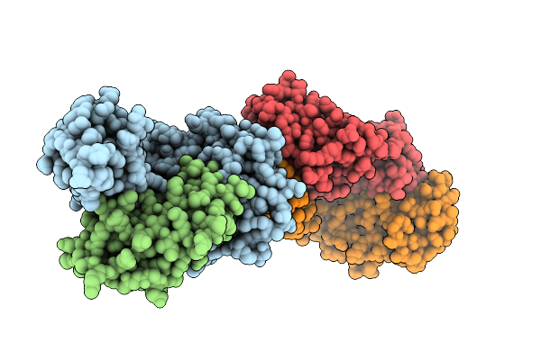 |
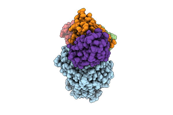
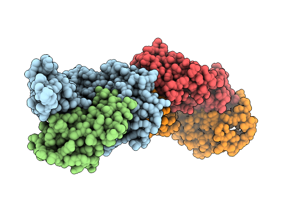
Organism: Homo sapiens, Mus musculus, Hepatitis b virus
Method: X-RAY DIFFRACTION Release Date: 2025-10-01 Classification: VIRAL PROTEIN/IMMUNE SYSTEM |
 |
Organism: Homo sapiens, Mus musculus, Hepatitis b virus
Method: X-RAY DIFFRACTION Release Date: 2025-10-01 Classification: VIRAL PROTEIN/IMMUNE SYSTEM |
 |
Organism: Homo sapiens, Mus musculus, Hepatitis b virus
Method: X-RAY DIFFRACTION Release Date: 2025-10-01 Classification: VIRAL PROTEIN/IMMUNE SYSTEM |
 |
Organism: Homo sapiens, Lama glama, Mus musculus
Method: ELECTRON MICROSCOPY Release Date: 2025-10-01 Classification: PEPTIDE BINDING PROTEIN/IMMUNE SYSTEM |