
Search Count: 1,394
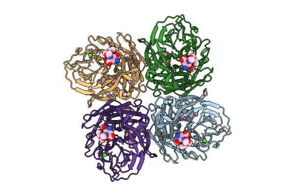 |
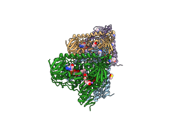
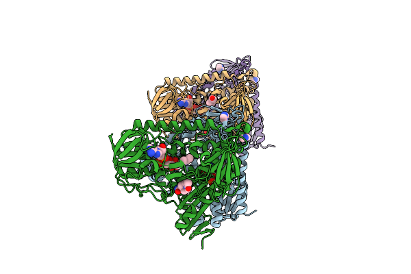
Organism: Influenza a virus
Method: ELECTRON MICROSCOPY Release Date: 2025-12-17 Classification: VIRAL PROTEIN Ligands: A1IVV, NAG, CA |
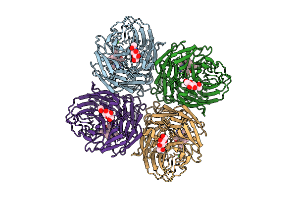 |
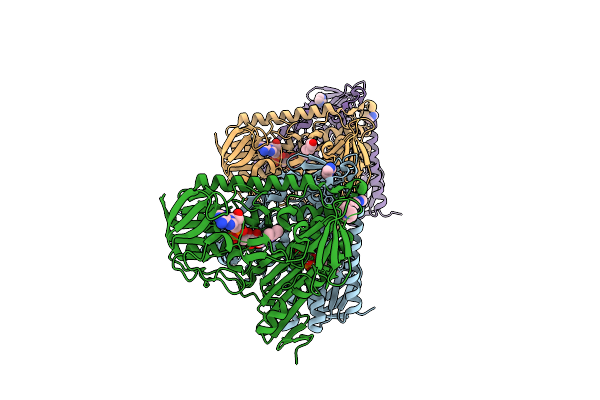
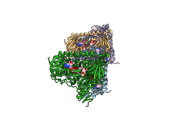
Organism: Influenza a virus
Method: ELECTRON MICROSCOPY Release Date: 2025-12-17 Classification: VIRAL PROTEIN Ligands: A1IVW, NAG, CA |
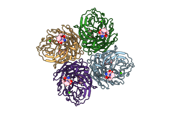 |
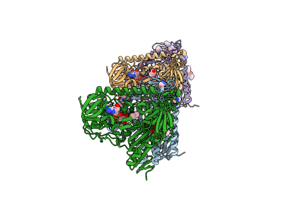
Organism: Influenza a virus
Method: ELECTRON MICROSCOPY Release Date: 2025-12-17 Classification: VIRAL PROTEIN Ligands: A1IVX, NAG, CA |
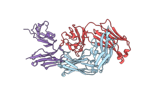 |
Organism: Homo sapiens
Method: X-RAY DIFFRACTION Release Date: 2025-12-17 Classification: IMMUNE SYSTEM |
 |
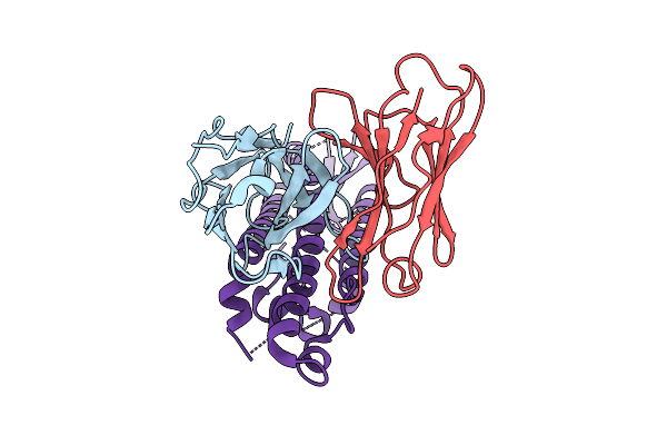
Conformational Flexibility In Hla-B8: Peptide Tuning Structural And Dynamical Changes
Organism: Homo sapiens, Human immunodeficiency virus 1
Method: X-RAY DIFFRACTION Release Date: 2025-12-10 Classification: IMMUNE SYSTEM |
 |
Conformational Flexibility In Hla-B8: Peptide Tuning Structural And Dynamical Changes
Organism: Homo sapiens, Human immunodeficiency virus 1
Method: X-RAY DIFFRACTION Release Date: 2025-12-10 Classification: IMMUNE SYSTEM |
 |
Conformational Flexibility In Hla-B8: Peptide Tuning Structural And Dynamical Changes
Organism: Homo sapiens, Human immunodeficiency virus 1
Method: X-RAY DIFFRACTION Release Date: 2025-12-10 Classification: IMMUNE SYSTEM |
 |
Nanoparticle Crystal Structure Of A Thermostabilized Mutant Rv1498A Flavoprotein From Mycobacterium Tuberculosis
Organism: Mycobacterium tuberculosis
Method: X-RAY DIFFRACTION Release Date: 2025-11-26 Classification: FLAVOPROTEIN |
 |
Nanoparticle Crystal Structure Of A Thermostabilized Mutant Of An As-Isolated Ftna (Ferritin) From Pseudomonas Aeruginosa
Organism: Pseudomonas aeruginosa
Method: X-RAY DIFFRACTION Release Date: 2025-11-26 Classification: METAL BINDING PROTEIN |
 |
Nanoparticle Crystal Structure Of A Thermostabilized Mutant E.Coli Ferritin Ecftna
Organism: Escherichia coli
Method: X-RAY DIFFRACTION Release Date: 2025-11-26 Classification: FLAVOPROTEIN |
 |
Organism: Homo sapiens, Severe acute respiratory syndrome coronavirus 2
Method: ELECTRON MICROSCOPY Release Date: 2025-11-26 Classification: VIRUS Ligands: NAG |
 |
Organism: Homo sapiens, Severe acute respiratory syndrome coronavirus 2
Method: ELECTRON MICROSCOPY Release Date: 2025-11-26 Classification: VIRUS |
 |
Pandda Analysis - Crystal Structure Of Trypanosoma Brucei Trypanothione Reductase In Complex With Z943693514
Organism: Trypanosoma brucei
Method: X-RAY DIFFRACTION Release Date: 2025-11-12 Classification: OXIDOREDUCTASE Ligands: FAD, IMD, PEG, DMS, BR, NA, GT4 |
 |
Pandda Analysis - Crystal Structure Of Trypanosoma Brucei Trypanothione Reductase In Complex With Z2856434836
Organism: Trypanosoma brucei
Method: X-RAY DIFFRACTION Release Date: 2025-11-12 Classification: OXIDOREDUCTASE Ligands: FAD, IMD, PEG, BR, LPZ |
 |
Pandda Analysis - Crystal Structure Of Trypanosoma Brucei Trypanothione Reductase In Complex With Z2204875953
Organism: Trypanosoma brucei
Method: X-RAY DIFFRACTION Release Date: 2025-11-12 Classification: OXIDOREDUCTASE Ligands: FAD, IMD, PEG, Q7L, BR |
 |
Pandda Analysis - Crystal Structure Of Trypanosoma Brucei Trypanothione Reductase In Complex With Z2856434890
Organism: Trypanosoma brucei
Method: X-RAY DIFFRACTION Release Date: 2025-11-12 Classification: OXIDOREDUCTASE Ligands: FAD, IMD, PEG, NZD, BR |
 |
Pandda Analysis - Crystal Structure Of Trypanosoma Brucei Trypanothione Reductase In Complex With Z32399802
Organism: Trypanosoma brucei
Method: X-RAY DIFFRACTION Release Date: 2025-11-12 Classification: OXIDOREDUCTASE Ligands: FAD, IMD, PEG, BR, A1I3U |
 |
Pandda Analysis - Crystal Structure Of Trypanosoma Brucei Trypanothione Reductase In Complex With Z2017861827
Organism: Trypanosoma brucei
Method: X-RAY DIFFRACTION Release Date: 2025-11-12 Classification: OXIDOREDUCTASE Ligands: FAD, IMD, PEG, A1BE9, BR |
 |
Pandda Analysis - Crystal Structure Of Trypanosoma Brucei Trypanothione Reductase In Complex With Z319545618
Organism: Trypanosoma brucei
Method: X-RAY DIFFRACTION Release Date: 2025-11-12 Classification: OXIDOREDUCTASE Ligands: FAD, IMD, PEG, RXS, BR, NA |
 |
Pandda Analysis - Crystal Structure Of Trypanosoma Brucei Trypanothione Reductase In Complex With Z436190540
Organism: Trypanosoma brucei
Method: X-RAY DIFFRACTION Release Date: 2025-11-12 Classification: OXIDOREDUCTASE Ligands: FAD, IMD, PEG, A1I3V, BR, NA |