
Search Count: 2,950
 |
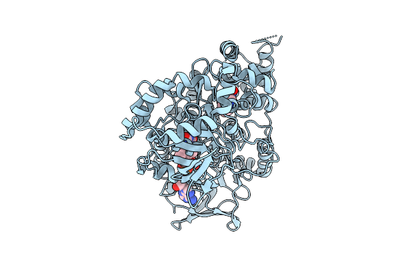
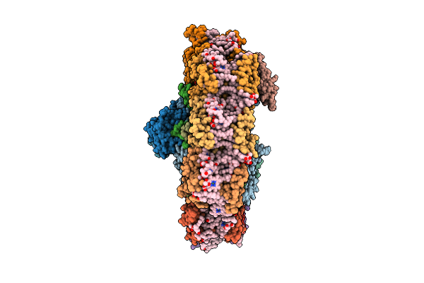
Organism: Human immunodeficiency virus 1, Homo sapiens
Method: ELECTRON MICROSCOPY Release Date: 2025-12-17 Classification: VIRAL PROTEIN/IMMUNE SYSTEM Ligands: NAG |
 |
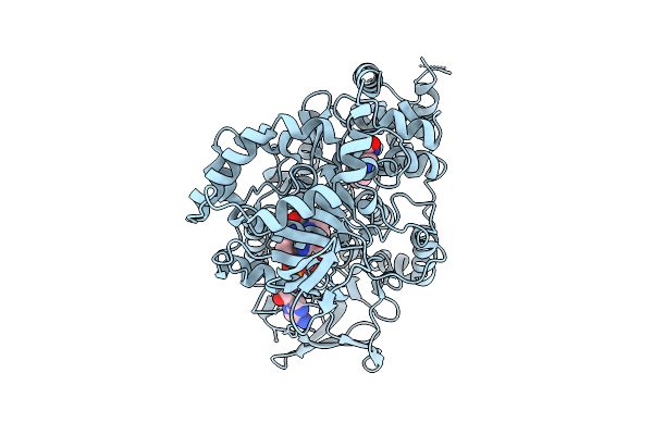
Organism: Human immunodeficiency virus 1, Homo sapiens
Method: ELECTRON MICROSCOPY Release Date: 2025-12-17 Classification: VIRAL PROTEIN/IMMUNE SYSTEM Ligands: NAG |
 |
Organism: Homo sapiens
Method: SOLUTION NMR Release Date: 2025-12-10 Classification: MEMBRANE PROTEIN |
 |
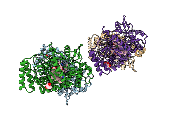
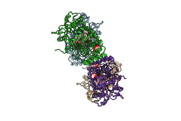
Organism: Triticum aestivum
Method: ELECTRON MICROSCOPY Release Date: 2025-12-10 Classification: PLANT PROTEIN |
 |
Organism: Aetokthonos hydrillicola thurmond2011
Method: X-RAY DIFFRACTION Release Date: 2025-12-10 Classification: FLAVOPROTEIN Ligands: FAD, TRP |
 |
Organism: Aetokthonos hydrillicola thurmond2011
Method: X-RAY DIFFRACTION Release Date: 2025-12-10 Classification: FLAVOPROTEIN Ligands: FAD, TRP |
 |
Organism: Aetokthonos hydrillicola thurmond2011
Method: X-RAY DIFFRACTION Release Date: 2025-12-10 Classification: FLAVOPROTEIN Ligands: FAD, TRP |
 |
Crystal Structure Of Aetf-L183F/V220I/S523A In Complex With Fad And L-Tryptophan
Organism: Aetokthonos hydrillicola thurmond2011
Method: X-RAY DIFFRACTION Release Date: 2025-12-10 Classification: FLAVOPROTEIN Ligands: TRP, FAD |
 |
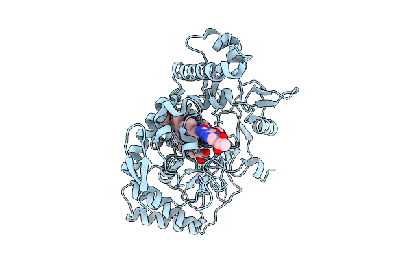
Structure Of Human Neuronal Nitric Oxide Synthase R354A/G357D Mutant Heme Domain Bound With 7-(3-Aminomethyl)Phenyl-6-Fluoro-4-Methylquinoin-2-Amine
Organism: Homo sapiens
Method: X-RAY DIFFRACTION Release Date: 2025-12-03 Classification: OXIDOREDUCTASE Ligands: HEM, H4B, A1BUD, GOL, ZN |
 |
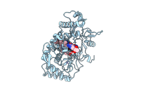
Structure Of Human Neuronal Nitric Oxide Synthase R354A/G357D Mutant Heme Domain Bound With 2-(2-Amino-6-Fluoro-4-Methylquinolin-7-Yl)-5-(Aminomethyl)Phenol
Organism: Homo sapiens
Method: X-RAY DIFFRACTION Release Date: 2025-12-03 Classification: OXIDOREDUCTASE Ligands: HEM, H4B, A1BUF, GOL, ZN |
 |
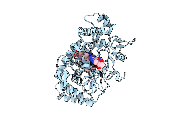
Structure Of Human Neuronal Nitric Oxide Synthase R354A/G357D Mutant Heme Domain Bound 2-(2-Amino-6-Fluoro-4-Methylquinolin-7-Yl)-5-(2-Aminoethyl)Phenol
Organism: Homo sapiens
Method: X-RAY DIFFRACTION Release Date: 2025-12-03 Classification: OXIDOREDUCTASE Ligands: HEM, H4B, A1BUH, GOL, ZN |
 |
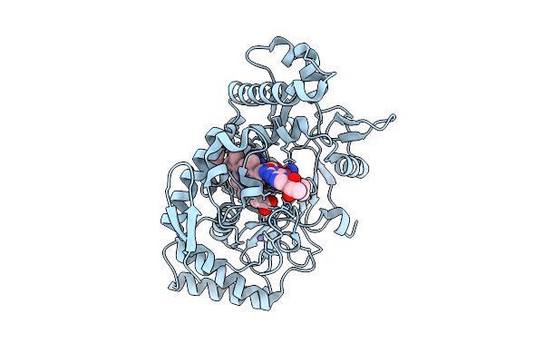
Structure Of Rat Neuronal Nitric Oxide Synthase R349A Mutant Heme Domain Bound With 7-(3-Aminomethyl)Phenyl-6-Fluoro-4-Methylquinoin-2-Amine
Organism: Rattus norvegicus
Method: X-RAY DIFFRACTION Release Date: 2025-12-03 Classification: OXIDOREDUCTASE Ligands: HEM, H4B, A1BUD, ACT, ZN |
 |
Structure Of Rat Neuronal Nitric Oxide Synthase R349A Mutant Heme Domain Bound With 2-(2-Amino-6-Fluoro-4-Methylquinolin-7-Yl)-5-(Aminomethyl)Phenol
Organism: Rattus norvegicus
Method: X-RAY DIFFRACTION Release Date: 2025-12-03 Classification: OXIDOREDUCTASE Ligands: HEM, H4B, ACT, ZN, A1BUF |
 |
Structure Of Rat Neuronal Nitric Oxide Synthase R349A Mutant Heme Domain Bound 2-(2-Amino-6-Fluoro-4-Methylquinolin-7-Yl)-5-(2-Aminoethyl)Phenol
Organism: Rattus norvegicus
Method: X-RAY DIFFRACTION Release Date: 2025-12-03 Classification: OXIDOREDUCTASE Ligands: HEM, H4B, ACT, ZN, A1BUH |
 |
Structure Of Rat Neuronal Nitric Oxide Synthase R349A Mutant Heme Domain Bound 7-(3-(Aminomethyl)-4-(Cyclopropylmethoxy)Phenyl)-6-Fluoro-4-Methylquinolin-2-Amine
Organism: Rattus norvegicus
Method: X-RAY DIFFRACTION Release Date: 2025-12-03 Classification: OXIDOREDUCTASE Ligands: HEM, H4B, A1BU3, ACT, ZN |
 |
Structure Of Human Endothelial Nitric Oxide Synthase Heme Domain Bound With 7-(3-Aminomethyl)Phenyl-6-Fluoro-4-Methylquinoin-2-Amine
Organism: Homo sapiens
Method: X-RAY DIFFRACTION Release Date: 2025-12-03 Classification: OXIDOREDUCTASE Ligands: HEM, H4B, A1BUD, BTB, GOL, CL, GD, ZN |
 |
Structure Of Human Endothelial Nitric Oxide Synthase Heme Domain Bound With 2-(2-Amino-6-Fluoro-4-Methylquinolin-7-Yl)-5-(Aminomethyl)Phenol
Organism: Homo sapiens
Method: X-RAY DIFFRACTION Release Date: 2025-12-03 Classification: OXIDOREDUCTASE Ligands: HEM, H4B, A1BUF, BTB, GOL, CL, GD, ZN, CA |
 |
Structure Of Human Endothelial Nitric Oxide Synthase Heme Domain Bound 2-(2-Amino-6-Fluoro-4-Methylquinolin-7-Yl)-5-(2-Aminoethyl)Phenol
Organism: Homo sapiens
Method: X-RAY DIFFRACTION Release Date: 2025-12-03 Classification: OXIDOREDUCTASE Ligands: HEM, H4B, A1BUH, BTB, ACT, GOL, CL, GD, ZN, CA |
 |
Structure Of Human Endothelial Nitric Oxide Synthase Heme Domain Bound 7-(3-(Aminomethyl)-4-(Cyclopropylmethoxy)Phenyl)-6-Fluoro-4-Methylquinolin-2-Amine
Organism: Homo sapiens
Method: X-RAY DIFFRACTION Release Date: 2025-12-03 Classification: OXIDOREDUCTASE Ligands: HEM, H4B, A1BU3, BTB, GOL, CL, GD, ZN, CA |
 |
Cryo-Em Structure Of Psi-Acpi From Rhodomonas Sp. Nies-2332 At 2.08 Angstroms Resolution
Organism: Rhodomonas sp. nies-2332
Method: ELECTRON MICROSCOPY Release Date: 2025-11-26 Classification: PHOTOSYNTHESIS Ligands: CL0, CLA, PQN, LHG, WVN, LMU, SF4, SQD, DGD, LMG, II0, IHT, KC2 |