Search Count: 316
 |
Organism: Homo sapiens
Method: X-RAY DIFFRACTION Release Date: 2025-06-25 Classification: STRUCTURAL PROTEIN Ligands: A1CF1, K |
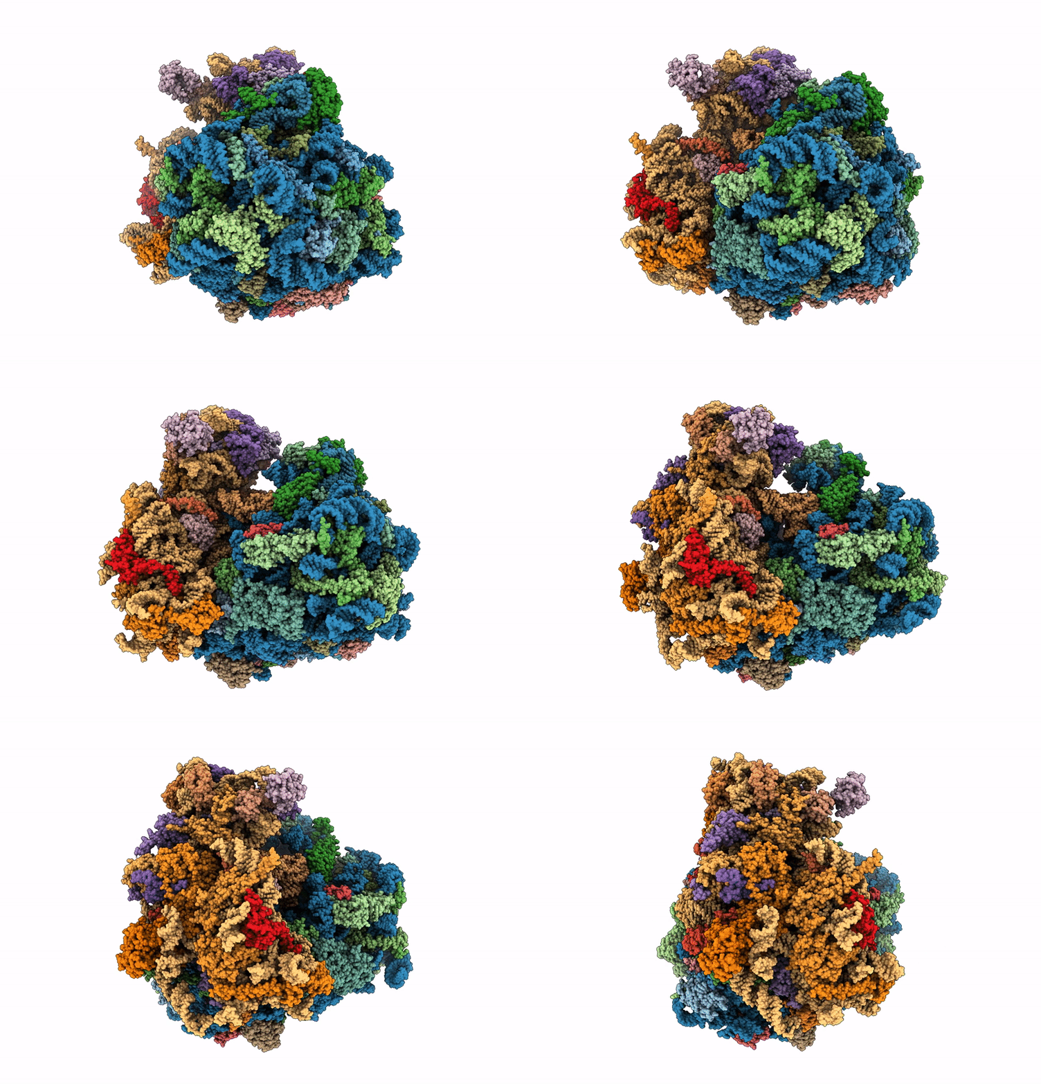 |
Single Particle Cryoem Structure Of The Pf80S Ribosome In Non-Rotated Pre State (Nrt A-P-E)
Organism: Plasmodium falciparum 3d7
Method: ELECTRON MICROSCOPY Release Date: 2025-05-28 Classification: RIBOSOME |
 |
Single Particle Cryoem Structure Of The Pf80S Ribosome In The Post State (Nrt With P- And E-Site Trna)
Organism: Plasmodium falciparum 3d7
Method: ELECTRON MICROSCOPY Release Date: 2025-05-28 Classification: RIBOSOME |
 |
Single Particle Cryoem Structure Of The Pf80S Ribosome In The Unloaded State (Nrt With E-Site Trna)
Organism: Plasmodium falciparum 3d7
Method: ELECTRON MICROSCOPY Release Date: 2025-05-28 Classification: RIBOSOME |
 |
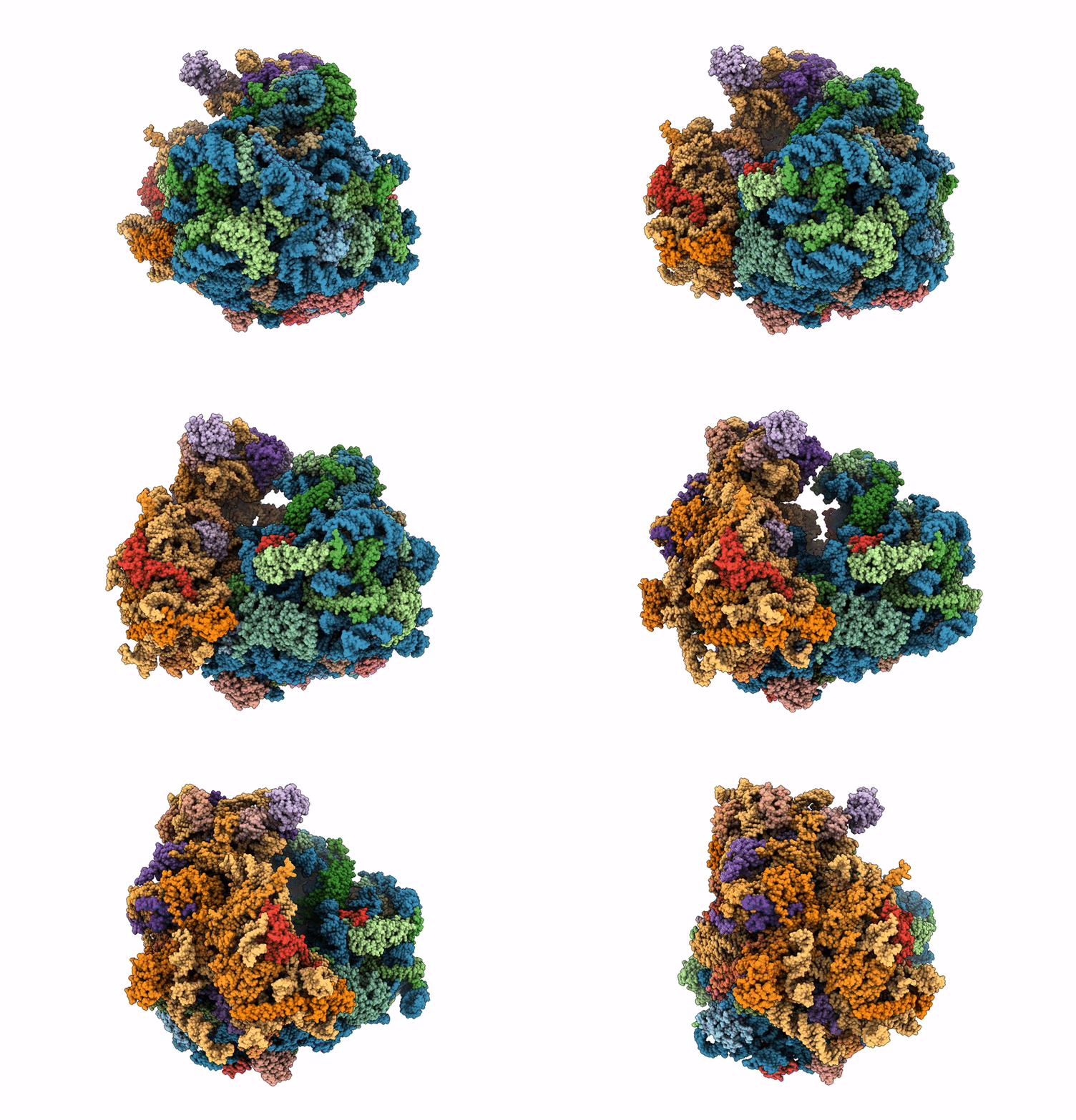
Single Particle Cryoem Structure Of The Pf80S Ribosome In The Rotated-2 Pre State (Rt State With P And E-Site Trna)
Organism: Plasmodium falciparum 3d7
Method: ELECTRON MICROSCOPY Release Date: 2025-05-28 Classification: RIBOSOME |
 |
Single Particle Cryoem Structure Of The Pf80S Ribosome In Rotated State With E-Site Trna
Organism: Plasmodium falciparum 3d7
Method: ELECTRON MICROSCOPY Release Date: 2025-05-28 Classification: RIBOSOME |
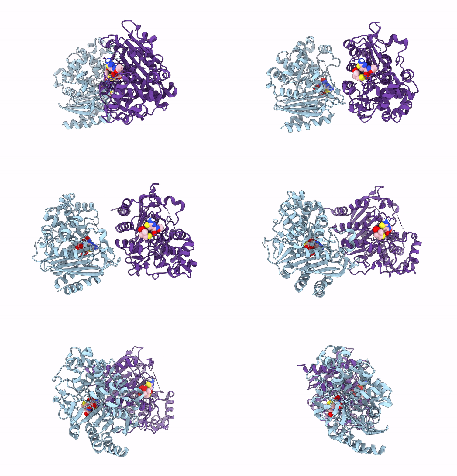 |
X-Ray Crystal Structure Of Adc-33 Beta-Lactamase In Complex With Ceftazidime In Acyl And Product Forms
Organism: Acinetobacter baumannii
Method: X-RAY DIFFRACTION Release Date: 2025-05-14 Classification: HYDROLASE Ligands: A1BIM, CAZ |
 |
Organism: Homo sapiens
Method: X-RAY DIFFRACTION Release Date: 2025-04-02 Classification: SIGNALING PROTEIN/HYDROLASE Ligands: GTP, MG |
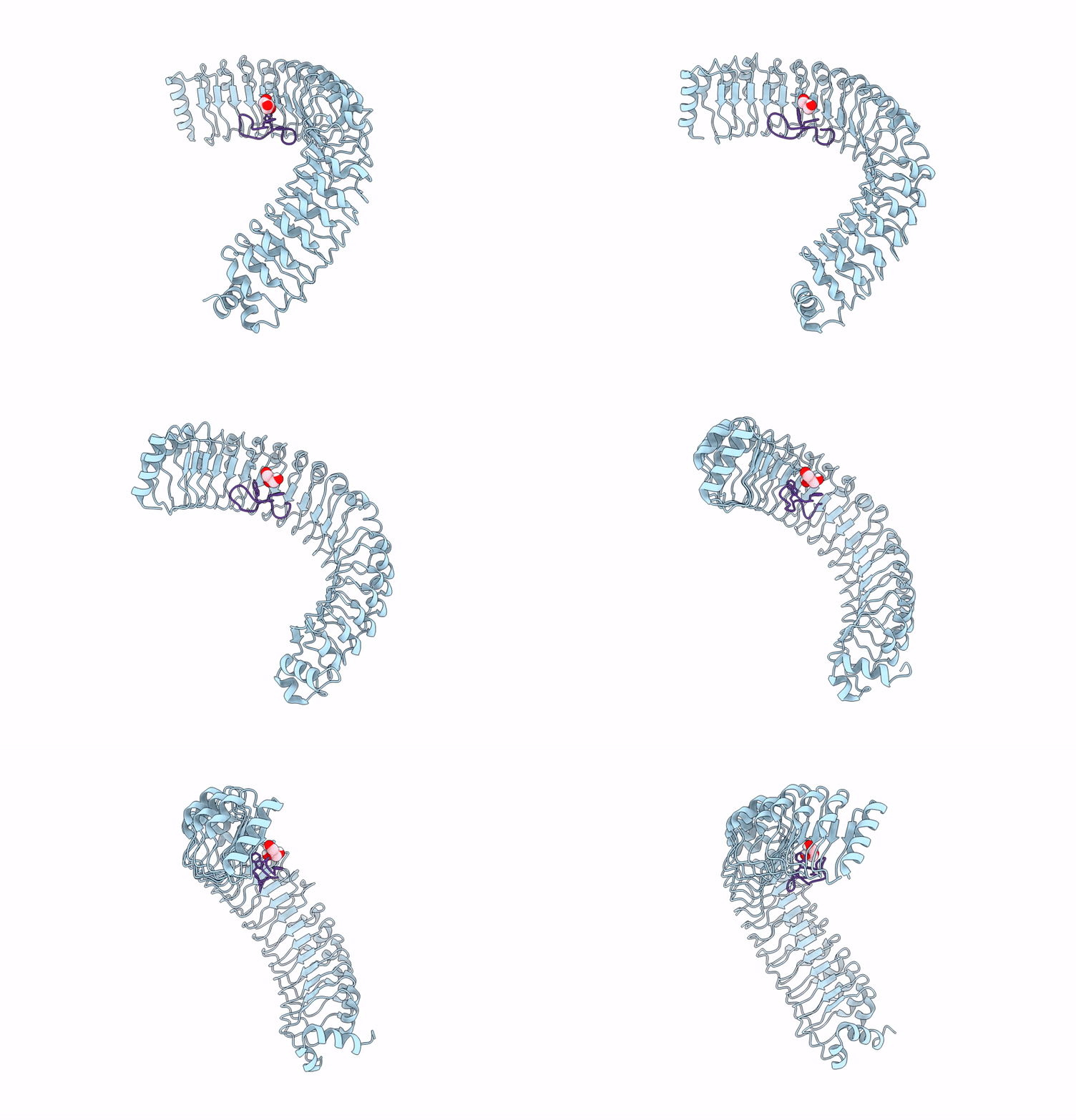 |
Organism: Homo sapiens, Synthetic construct
Method: X-RAY DIFFRACTION Release Date: 2025-04-02 Classification: STRUCTURAL PROTEIN Ligands: GOL |
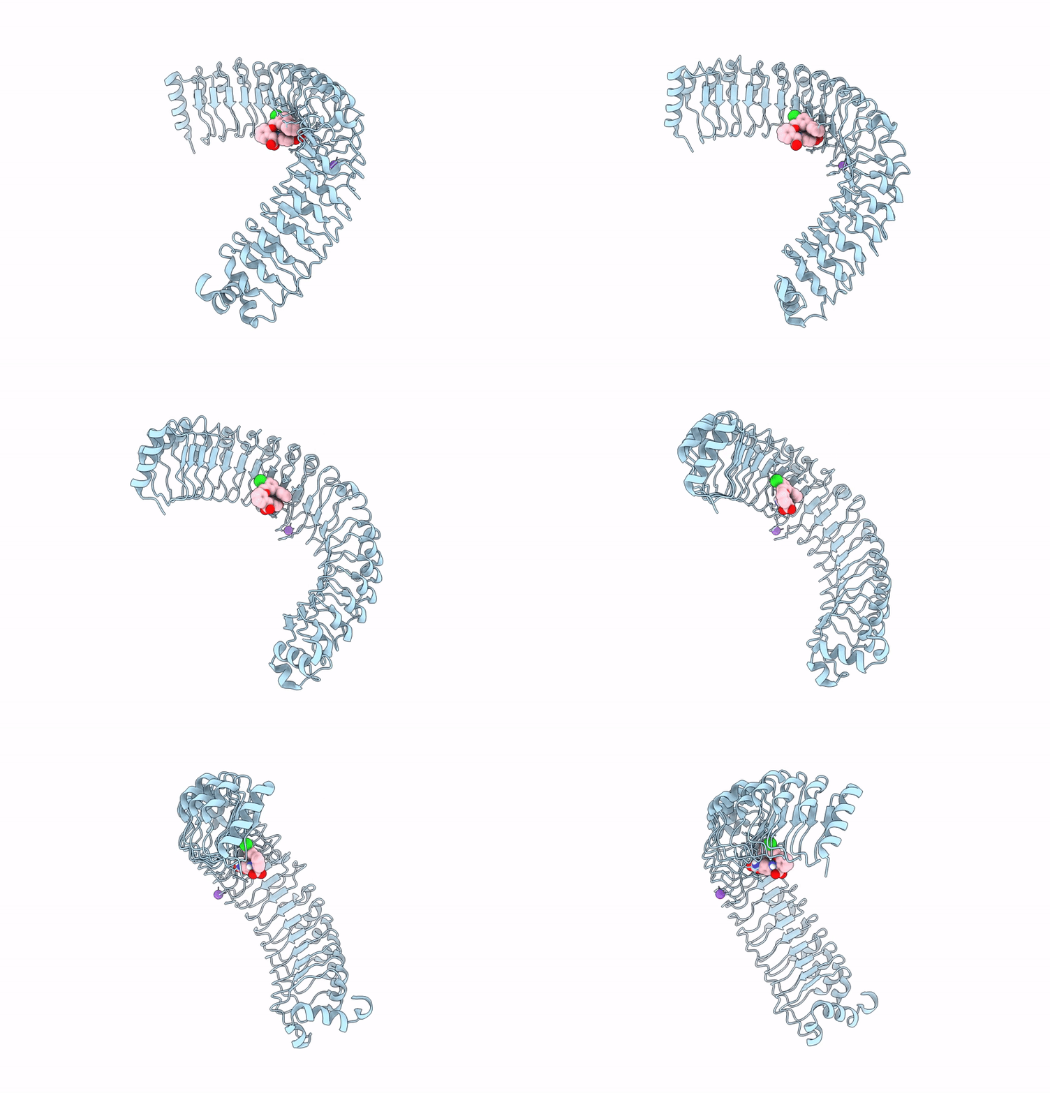 |
Organism: Homo sapiens
Method: X-RAY DIFFRACTION Release Date: 2025-04-02 Classification: STRUCTURAL PROTEIN Ligands: A1ATZ, K |
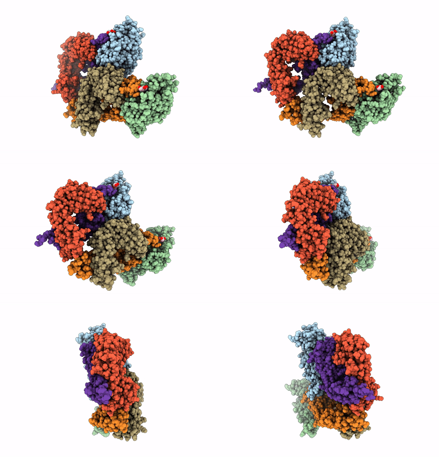 |
Organism: Severe acute respiratory syndrome coronavirus 2, Homo sapiens
Method: X-RAY DIFFRACTION Resolution:2.60 Å Release Date: 2025-03-26 Classification: VIRAL PROTEIN Ligands: NAG |
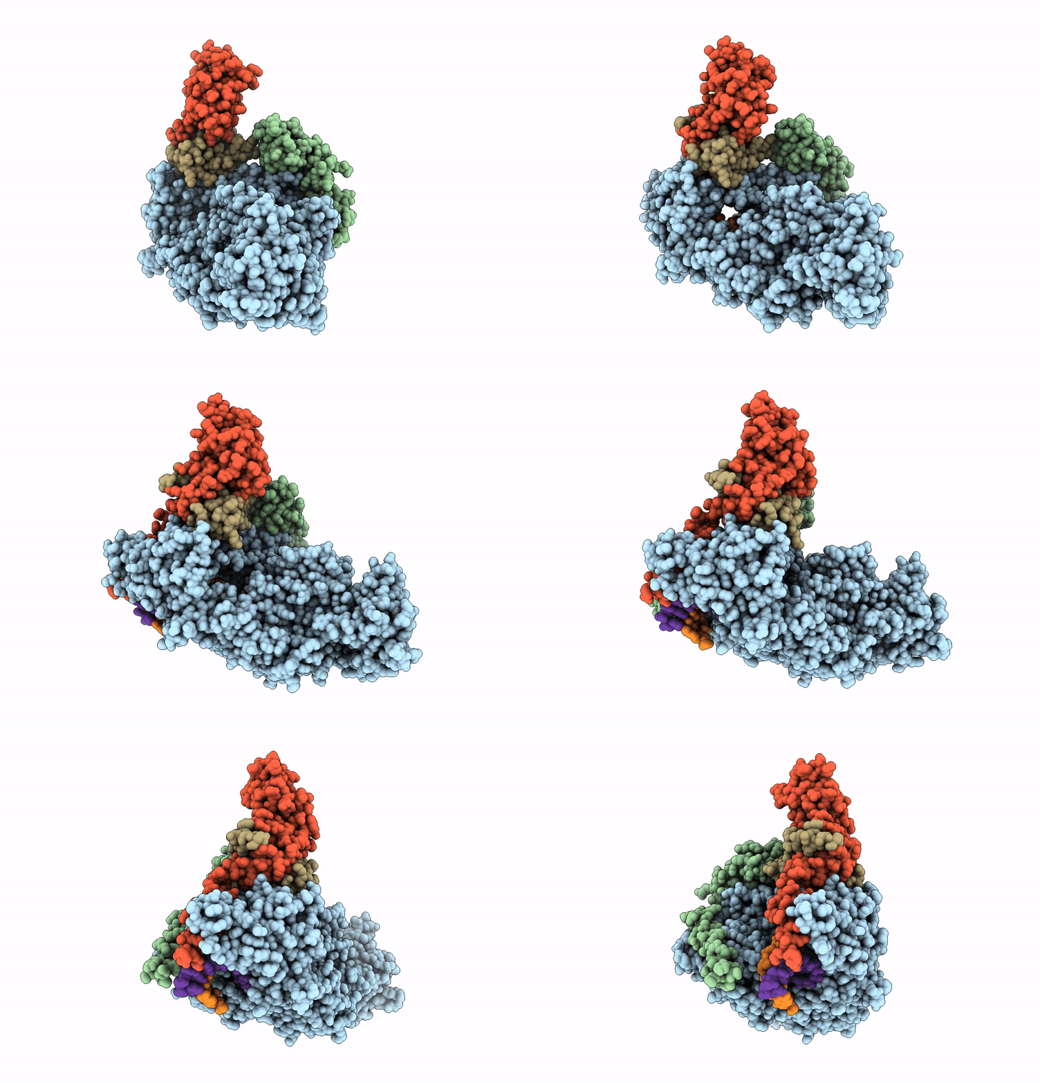 |
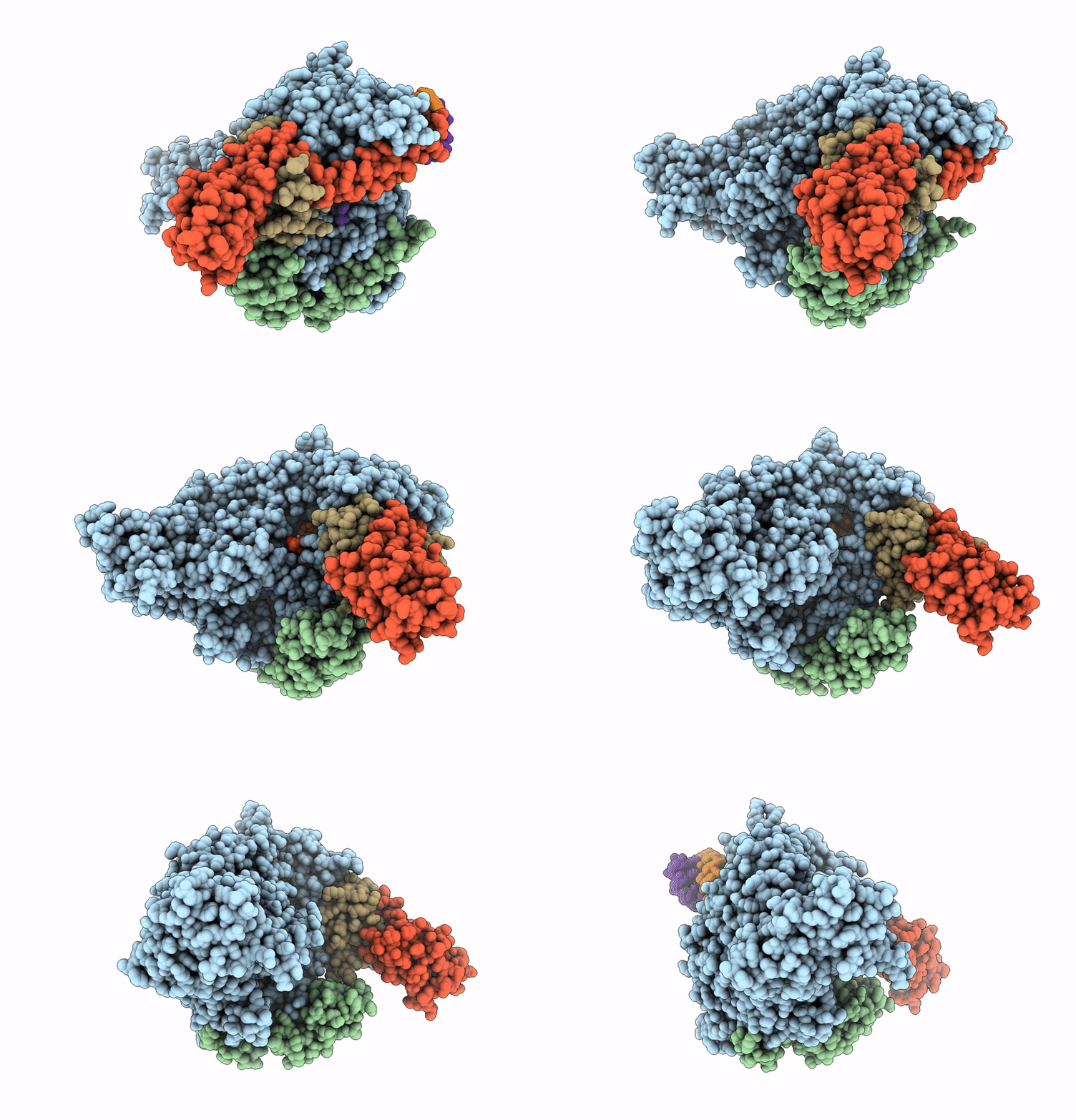
Organism: Severe acute respiratory syndrome coronavirus 2
Method: ELECTRON MICROSCOPY Resolution:2.90 Å Release Date: 2025-03-19 Classification: VIRAL PROTEIN/RNA Ligands: ZN, MG |
 |
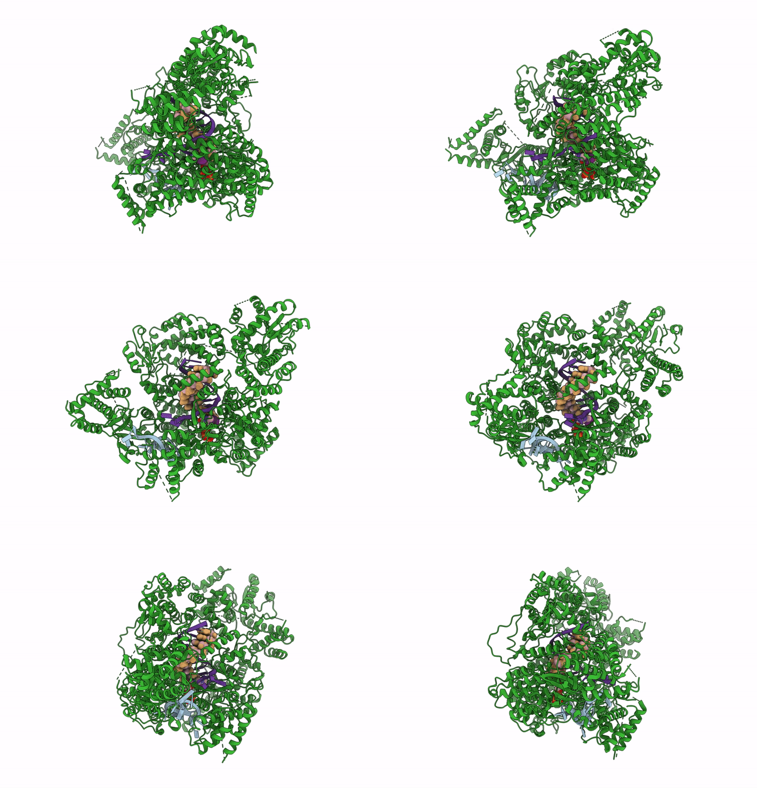
Cryoem Structure Of Compound Hnc-1664 Bound With Rdrp-Rna Complex Of Sars-Cov-2
Organism: Severe acute respiratory syndrome coronavirus 2, Severe acute respiratory syndrome coronavirus, Synthetic construct
Method: ELECTRON MICROSCOPY Release Date: 2024-12-04 Classification: VIRAL PROTEIN/RNA Ligands: A1LVZ, MG |
 |
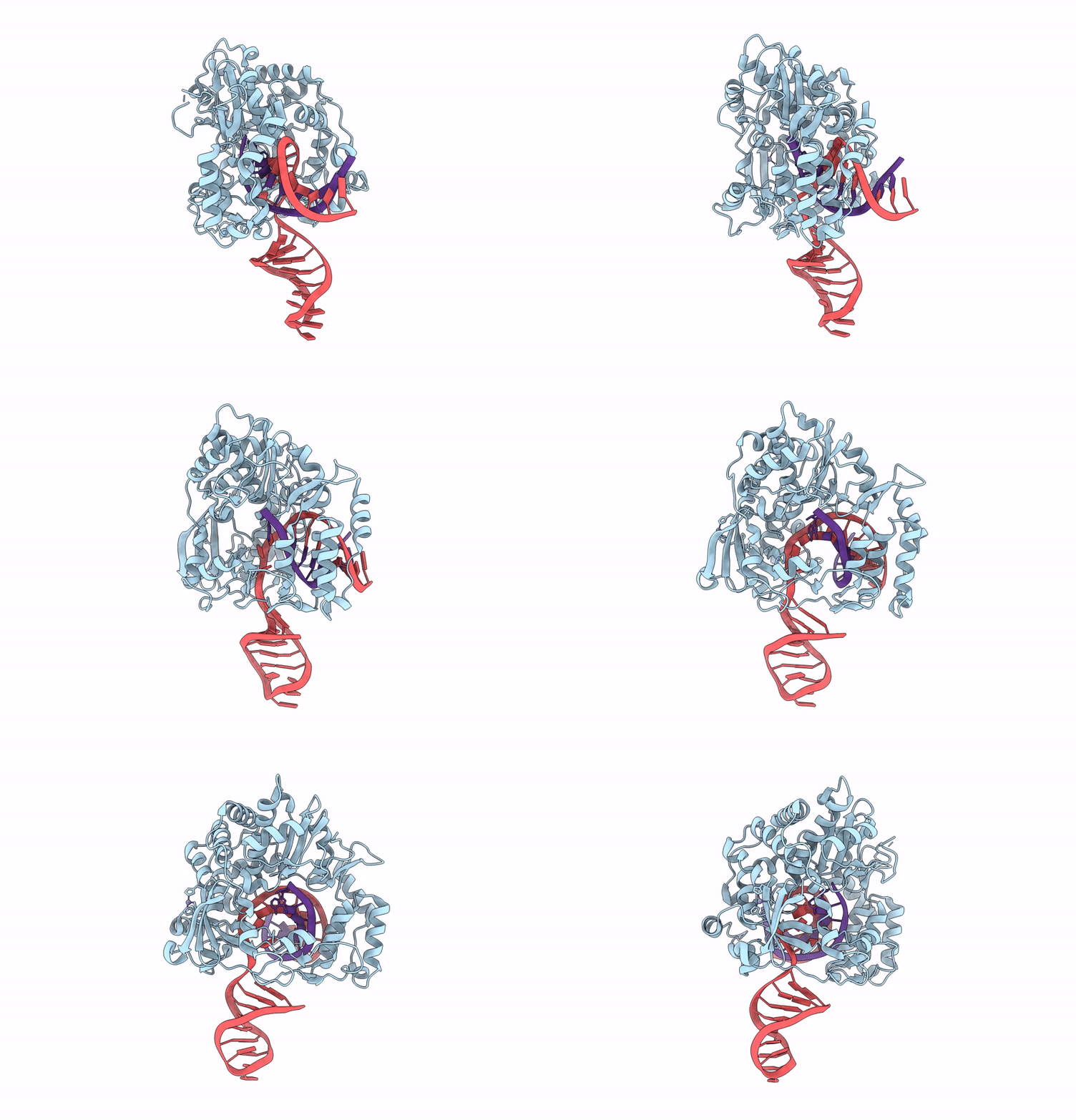
Cryo-Em Structure Of Lassa Virus Rdrp Elongation Complex With The Ntp Form Of Compound Hnc-1664 Bound In The Active Site
Organism: Lassa virus josiah, Synthetic construct
Method: ELECTRON MICROSCOPY Release Date: 2024-12-04 Classification: REPLICATION Ligands: A1LVZ |
 |
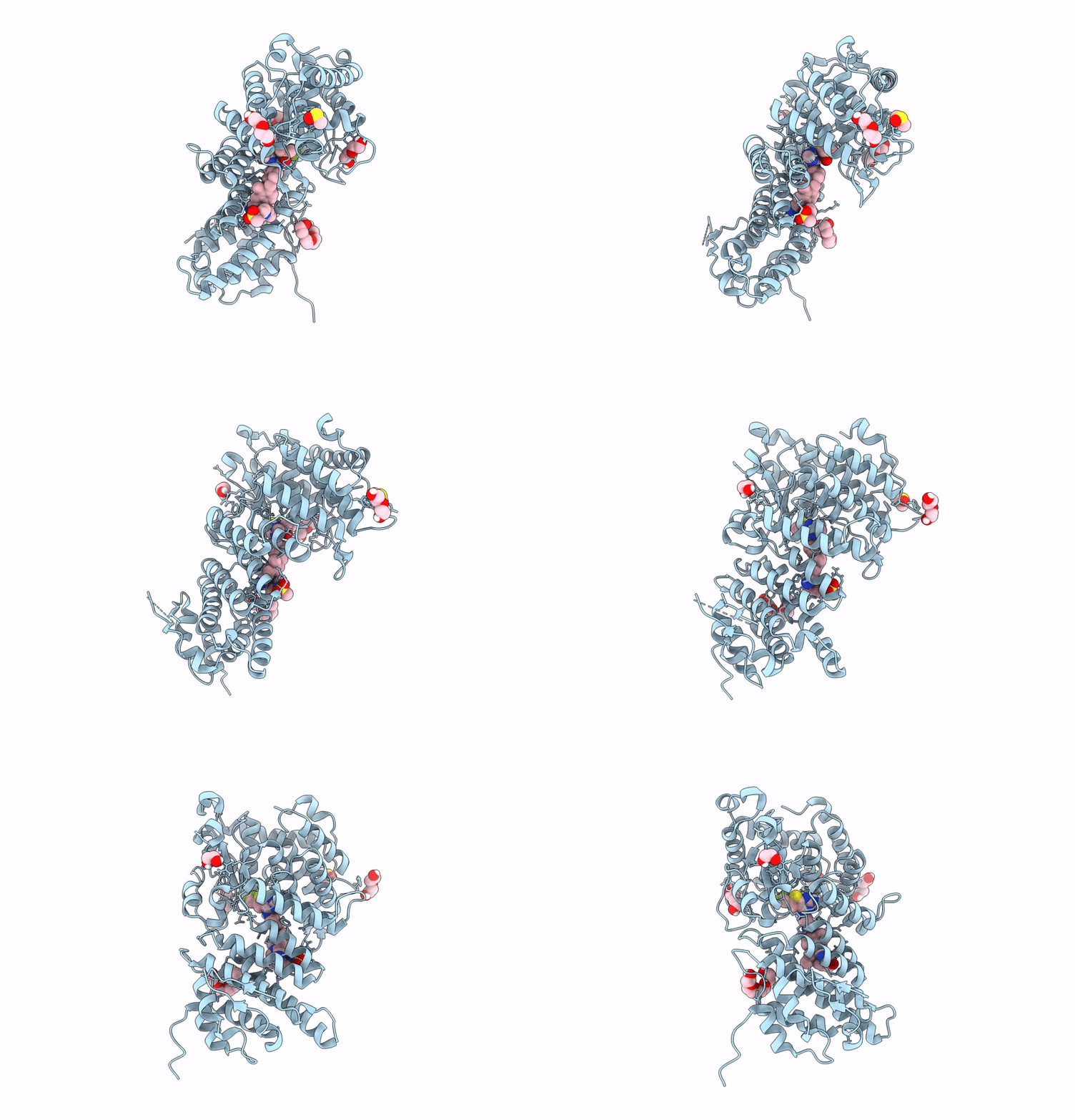
Crystal Structure Of The Enterovirus 71 Rdrp Elongation Complex With The Nucleoside Monophosphate Form Of Compound Hnc-1664 At Product Position -1 (Post-Translocation State)
Organism: Enterovirus a71, Synthetic construct
Method: X-RAY DIFFRACTION Resolution:3.00 Å Release Date: 2024-12-04 Classification: REPLICATION Ligands: ZN |
 |
Organism: Homo sapiens
Method: X-RAY DIFFRACTION Release Date: 2024-11-20 Classification: PROTEIN BINDING Ligands: K5O, PEG, PG4, EDO, DMS |
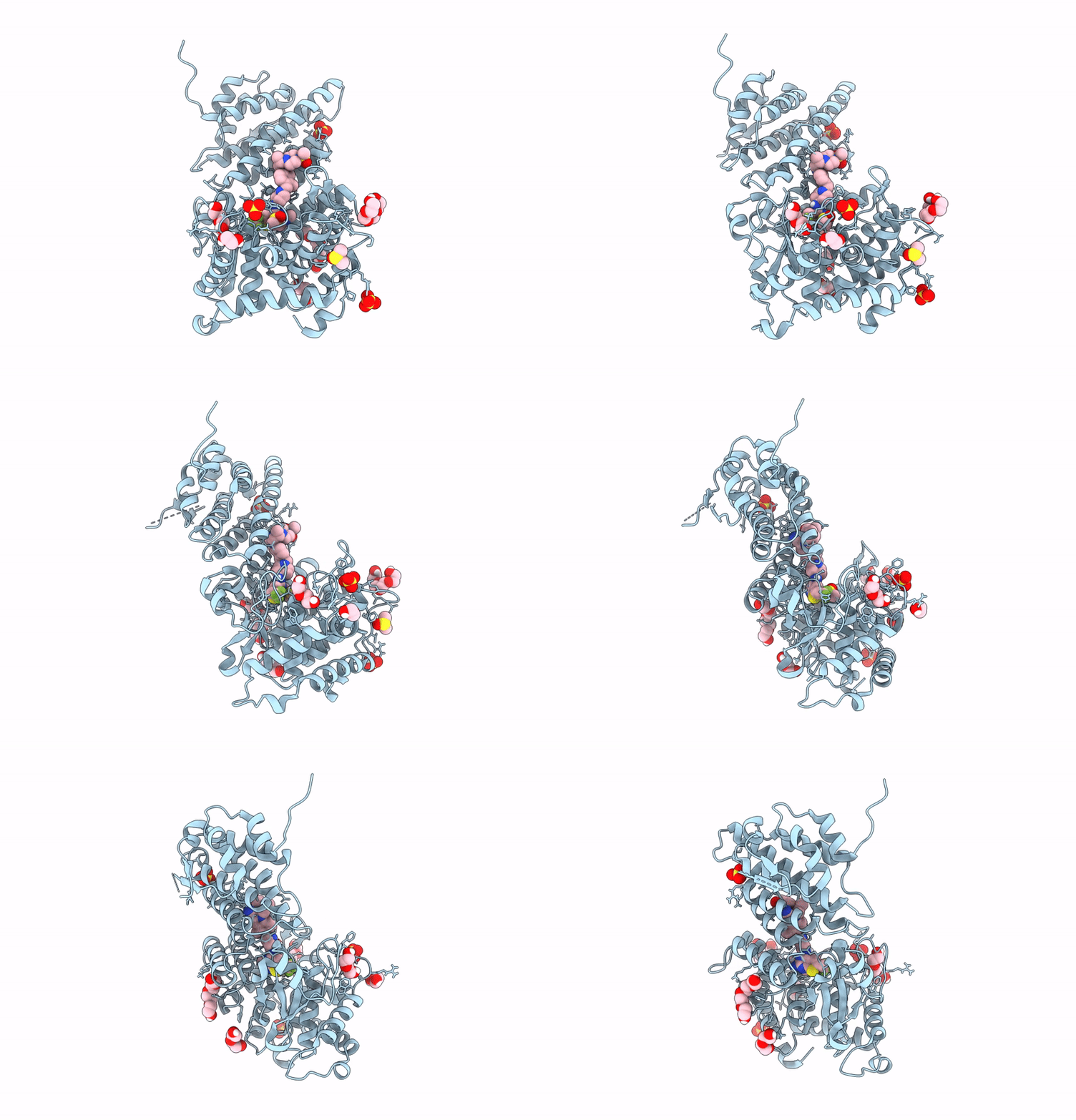 |
Organism: Homo sapiens
Method: X-RAY DIFFRACTION Release Date: 2024-11-20 Classification: PROTEIN BINDING Ligands: K5O, PEG, SO4, DMS, PG4, EDO |
 |
Crystal Structure Of Heparosan Synthase 2 From Pasteurella Multocida At 2.85 A
Organism: Pasteurella multocida
Method: X-RAY DIFFRACTION Resolution:2.85 Å Release Date: 2024-09-04 Classification: TRANSFERASE Ligands: MN, UDP, NA, CL, EDO, CA |
 |
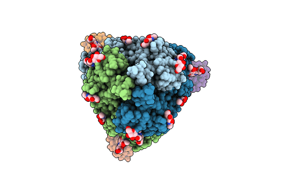
Subtomogram Averaged Consensus Structure Of The Malarial 80S Ribosome In Plasmodium Falciparum-Infected Human Erythrocytes
Organism: Plasmodium falciparum 3d7
Method: ELECTRON MICROSCOPY Release Date: 2024-08-14 Classification: RIBOSOME |
 |
Organism: Severe acute respiratory syndrome coronavirus 2, Homo sapiens
Method: ELECTRON MICROSCOPY Release Date: 2024-07-31 Classification: VIRAL PROTEIN Ligands: NAG |

