Planned Maintenance: Some services may turn out to be unavailable from 15th January, 2026 to 16th January, 2026. We apologize for the inconvenience!

Planned Maintenance: Some services may turn out to be unavailable from 15th January, 2026 to 16th January, 2026. We apologize for the inconvenience!
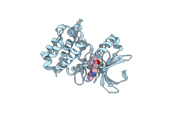 |
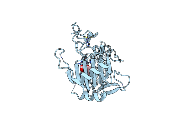
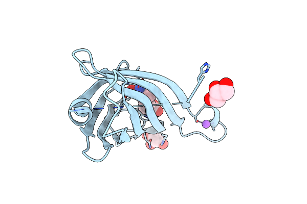
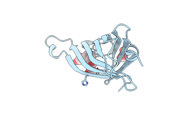
Organism: Homo sapiens
Method: X-RAY DIFFRACTION Resolution:1.80 Å Release Date: 2025-01-15 Classification: SIGNALING PROTEIN Ligands: OG0 |
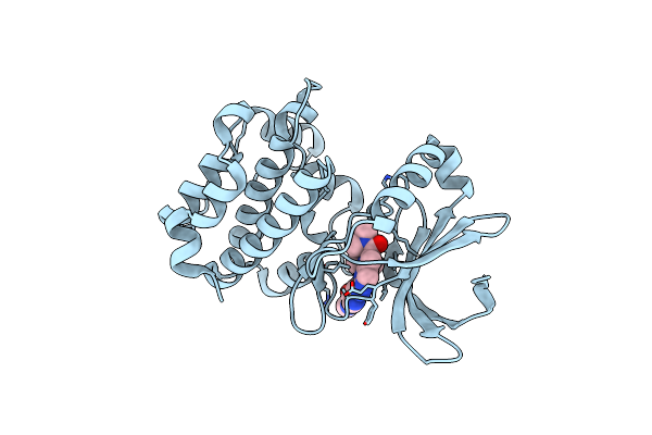 |
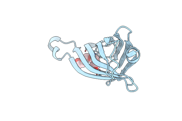
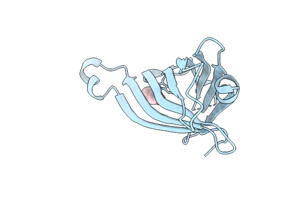
Organism: Homo sapiens
Method: X-RAY DIFFRACTION Resolution:1.81 Å Release Date: 2025-01-15 Classification: SIGNALING PROTEIN Ligands: OGR |
 |
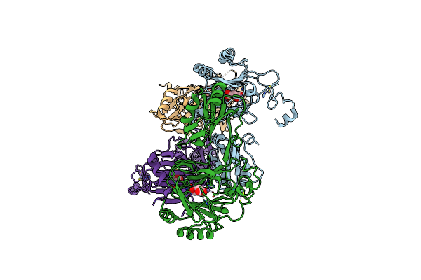
Organism: Thermococcus kodakarensis
Method: ELECTRON MICROSCOPY Release Date: 2020-07-29 Classification: RIBOSOME Ligands: ZN |
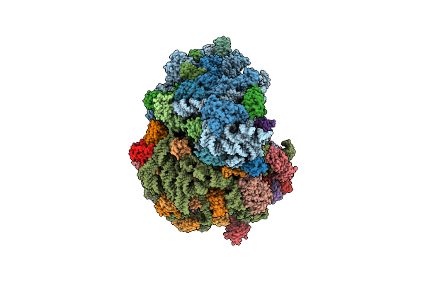 |
Cryo-Em Structure Of T. Kodakarensis 70S Ribosome In Tknat10 Deleted Strain
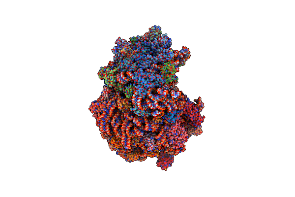
Organism: Thermococcus kodakarensis
Method: ELECTRON MICROSCOPY Release Date: 2020-07-29 Classification: RIBOSOME Ligands: ZN |
 |
Organism: Thermococcus kodakarensis (strain atcc baa-918 / jcm 12380 / kod1)
Method: ELECTRON MICROSCOPY Release Date: 2020-07-29 Classification: RIBOSOME Ligands: ZN |
 |
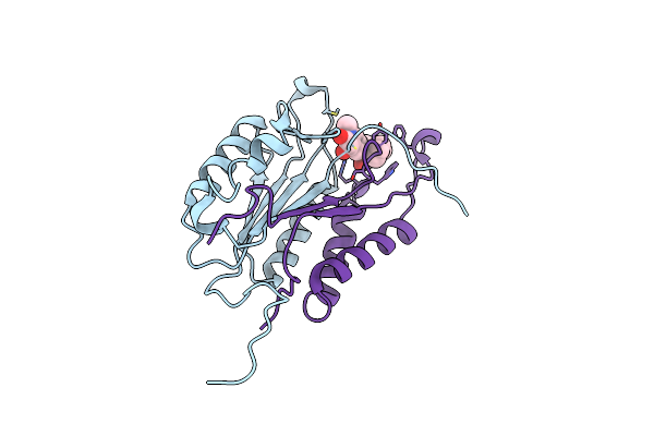
Crystal Structure And Rna Binding Properties Of The Rrm/Alkb Domains In Human Abh8, An Enzyme Catalyzing Trna Hypermodification, Northeast Structural Genomics Consortium Target Hr5601B
Organism: Homo sapiens
Method: X-RAY DIFFRACTION Resolution:3.20 Å Release Date: 2011-11-02 Classification: OXIDOREDUCTASE Ligands: ZN, MN, AKG |
 |
Crystal Structure And Rna Binding Properties Of The Rrm/Alkb Domains In Human Abh8, An Enzyme Catalyzing Trna Hypermodification, Northeast Structural Genomics Consortium Target Hr5601B
Organism: Homo sapiens
Method: X-RAY DIFFRACTION Resolution:3.01 Å Release Date: 2011-11-02 Classification: OXIDOREDUCTASE Ligands: ZN, MN, AKG |
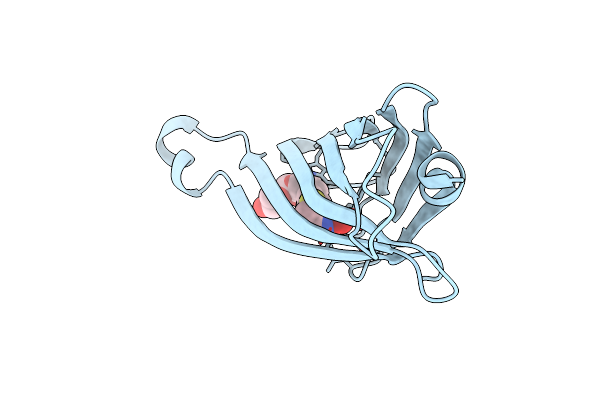 |
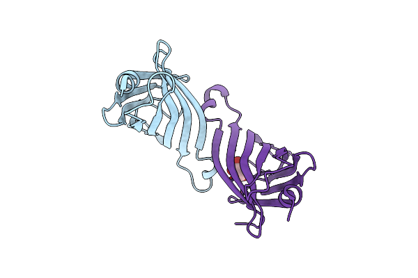
Organism: Streptomyces avidinii
Method: X-RAY DIFFRACTION Resolution:1.60 Å Release Date: 2011-07-06 Classification: BIOTIN BINDING PROTEIN Ligands: 1PE, BTN |
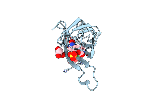 |
Organism: Streptomyces avidinii
Method: X-RAY DIFFRACTION Resolution:1.40 Å Release Date: 2011-07-06 Classification: BIOTIN BINDING PROTEIN Ligands: BTN, NI, SO4, GOL |
 |
Organism: Streptomyces avidinii
Method: X-RAY DIFFRACTION Resolution:1.60 Å Release Date: 2011-07-06 Classification: BIOTIN BINDING PROTEIN Ligands: DTB, NI, NA, GOL |
 |
Organism: Streptomyces avidinii
Method: X-RAY DIFFRACTION Resolution:1.50 Å Release Date: 2011-07-06 Classification: BIOTIN BINDING PROTEIN Ligands: 1PE |
 |
Organism: Streptomyces avidinii
Method: X-RAY DIFFRACTION Resolution:1.50 Å Release Date: 2011-07-06 Classification: BIOTIN BINDING PROTEIN Ligands: GOL, 1PE |
 |
Organism: Streptomyces avidinii
Method: X-RAY DIFFRACTION Resolution:2.10 Å Release Date: 2011-07-06 Classification: BIOTIN BINDING PROTEIN Ligands: GOL |
 |
Organism: Streptomyces avidinii
Method: X-RAY DIFFRACTION Resolution:1.95 Å Release Date: 2011-07-06 Classification: BIOTIN BINDING PROTEIN Ligands: 1PE, GOL |
 |
Organism: Streptomyces avidinii
Method: X-RAY DIFFRACTION Resolution:1.82 Å Release Date: 2011-07-06 Classification: BIOTIN BINDING PROTEIN Ligands: GOL |
 |
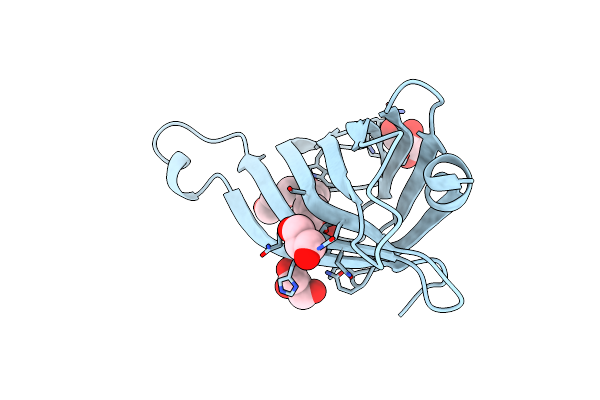
Organism: Homo sapiens
Method: X-RAY DIFFRACTION Resolution:2.75 Å Release Date: 2010-02-09 Classification: RNA BINDING PROTEIN Ligands: GOL |
 |
Crystal Structure Of The Cysteine Protease Human Cathepsin K In Complex With A Covalent Azepanone Inhibitor
Organism: Homo sapiens
Method: X-RAY DIFFRACTION Resolution:2.80 Å Release Date: 2003-01-14 Classification: HYDROLASE Ligands: 750 |
 |
Crystal Structure Of The Cysteine Protease Human Cathepsin K In Complex With A Covalent Azepanone Inhibitor
Organism: Homo sapiens
Method: X-RAY DIFFRACTION Resolution:2.40 Å Release Date: 2003-01-14 Classification: HYDROLASE Ligands: 2CA |
 |
The 2.8 Angstrom Crystal Structure Of Caspase-3 (Apopain Or Cpp32)In Complex With An Isatin Sulfonamide Inhibitor.
Organism: Homo sapiens
Method: X-RAY DIFFRACTION Resolution:2.80 Å Release Date: 2000-06-23 Classification: HYDROLASE Ligands: MSI |
 |
Crystal Structure Of Cysteine Protease Human Cathepsin K In Complex With A Covalent Peptidomimetic Inhibitor
Organism: Homo sapiens
Method: X-RAY DIFFRACTION Resolution:2.30 Å Release Date: 1999-06-08 Classification: HYDROLASE Ligands: I10 |