
Search Count: 12
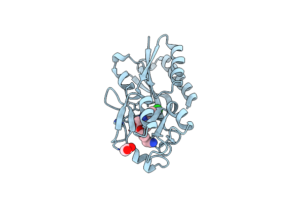 |
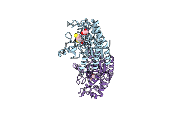
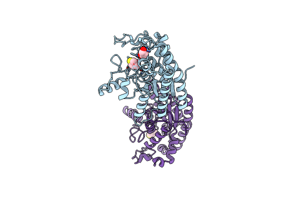
Pqsr Coinducer Binding Domain Of Pseudomonas Aeruginosa With Ligand 2T : 2-(4-(3-((6-Chloro-1-(2-Methoxyethyl)-1H-Benzo[D]Imidazol-2-Yl)Amino)-2-Hydroxypropoxy)Phenyl)Acetonitrile
Organism: Pseudomonas aeruginosa ucbpp-pa14
Method: X-RAY DIFFRACTION Resolution:2.80 Å Release Date: 2024-01-17 Classification: TRANSCRIPTION Ligands: JTI, EDO |
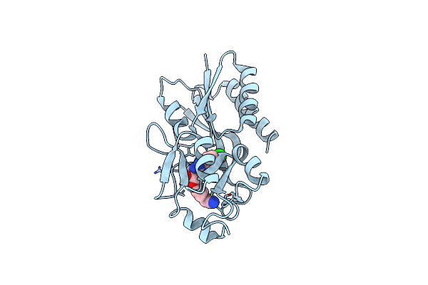 |
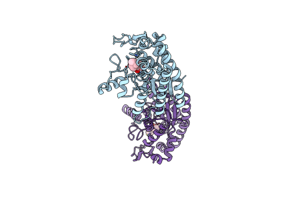
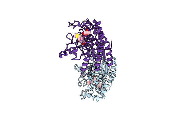
Pqsr Coinducer Binding Domain Of Pseudomonas Aeruginosa With Ligand 2F: 2-(4-(3-((6-Chloro-1-Isopropyl-1H-Benzo[D]Imidazol-2-Yl)Amino)-2-Hydroxypropoxy)Phenyl)Acetonitrile
Organism: Pseudomonas aeruginosa ucbpp-pa14
Method: X-RAY DIFFRACTION Resolution:2.90 Å Release Date: 2024-01-17 Classification: TRANSCRIPTION Ligands: JVO |
 |
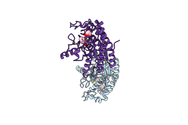
Organism: Pseudomonas aeruginosa ucbpp-pa14
Method: X-RAY DIFFRACTION Resolution:3.20 Å Release Date: 2020-05-13 Classification: DNA BINDING PROTEIN Ligands: NV5 |
 |
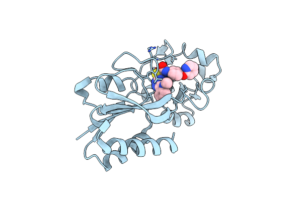
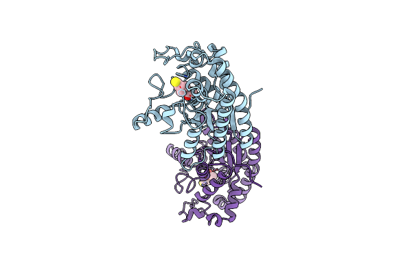
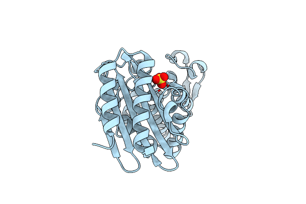
Crystal Structure Of Choe, A Bacterial Acetylcholinesterase From Pseudomonas Aeruginosa
Organism: Pseudomonas aeruginosa (strain atcc 15692 / dsm 22644 / cip 104116 / jcm 14847 / lmg 12228 / 1c / prs 101 / pao1)
Method: X-RAY DIFFRACTION Resolution:1.35 Å Release Date: 2020-05-13 Classification: HYDROLASE Ligands: MES, GOL, BUA, CL, P6G |
 |
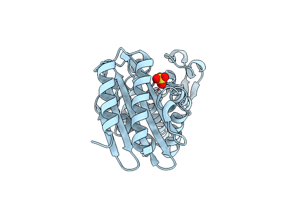
Organism: Pseudomonas aeruginosa (strain atcc 15692 / dsm 22644 / cip 104116 / jcm 14847 / lmg 12228 / 1c / prs 101 / pao1)
Method: X-RAY DIFFRACTION Resolution:1.65 Å Release Date: 2020-05-13 Classification: HYDROLASE Ligands: ACT, ETM, GOL |
 |
Organism: Pseudomonas aeruginosa (strain atcc 15692 / dsm 22644 / cip 104116 / jcm 14847 / lmg 12228 / 1c / prs 101 / pao1)
Method: X-RAY DIFFRACTION Resolution:1.85 Å Release Date: 2020-05-13 Classification: HYDROLASE Ligands: QFJ, IOD, PPI, ETM |
 |
Organism: Pseudomonas aeruginosa (strain atcc 15692 / dsm 22644 / cip 104116 / jcm 14847 / lmg 12228 / 1c / prs 101 / pao1)
Method: X-RAY DIFFRACTION Resolution:1.57 Å Release Date: 2020-05-13 Classification: HYDROLASE Ligands: AT3, GOL |
 |
Crystal Structure Of Choe D285N Mutant In Complex With Acetate And Thiocholine
Organism: Pseudomonas aeruginosa (strain atcc 15692 / dsm 22644 / cip 104116 / jcm 14847 / lmg 12228 / 1c / prs 101 / pao1)
Method: X-RAY DIFFRACTION Resolution:1.43 Å Release Date: 2020-05-13 Classification: HYDROLASE Ligands: ACT, ETM |
 |
Organism: Pseudomonas aeruginosa (strain atcc 15692 / dsm 22644 / cip 104116 / jcm 14847 / lmg 12228 / 1c / prs 101 / pao1)
Method: X-RAY DIFFRACTION Resolution:1.80 Å Release Date: 2020-05-13 Classification: HYDROLASE Ligands: ETM, GOL |
 |
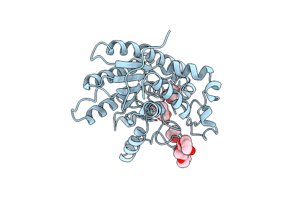
Crystal Structure Of Choe S38A Mutant In Complex With Acetate And Acetylthiocholine
Organism: Pseudomonas aeruginosa (strain atcc 15692 / dsm 22644 / cip 104116 / jcm 14847 / lmg 12228 / 1c / prs 101 / pao1)
Method: X-RAY DIFFRACTION Resolution:1.42 Å Release Date: 2020-05-13 Classification: HYDROLASE Ligands: ACT, AT3, GOL |
 |
Organism: Pseudomonas aeruginosa
Method: X-RAY DIFFRACTION Resolution:1.95 Å Release Date: 2001-02-21 Classification: HYDROLASE Ligands: SO4 |
 |
Organism: Pseudomonas aeruginosa
Method: X-RAY DIFFRACTION Resolution:1.75 Å Release Date: 2001-02-21 Classification: HYDROLASE Ligands: SO4 |