
Search Count: 46
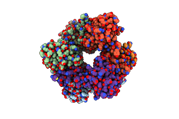 |
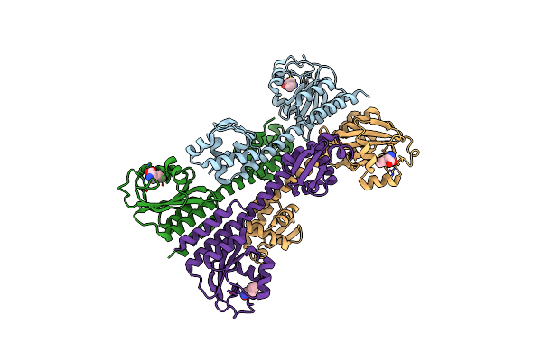
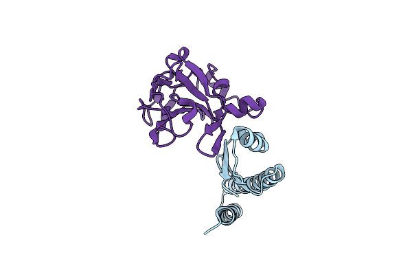
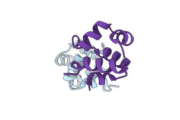
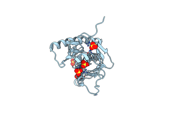
The Structure Of The Cofactor Binding Gaf Domain Of The Nutrient Sensor Cody From Clostridium Difficile
Organism: Clostridioides difficile
Method: X-RAY DIFFRACTION Resolution:1.68 Å Release Date: 2018-02-14 Classification: TRANSCRIPTION Ligands: ILE |
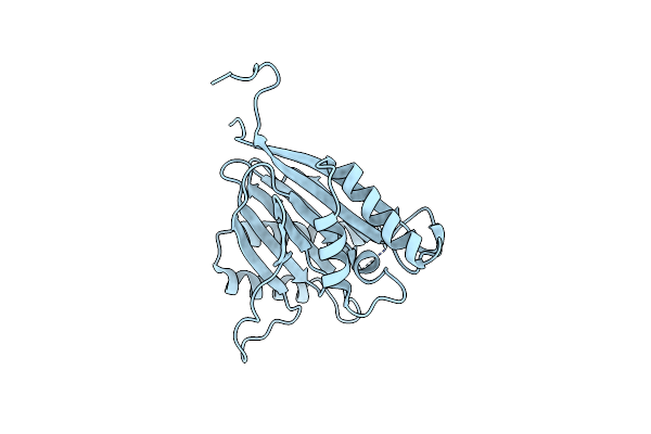 |
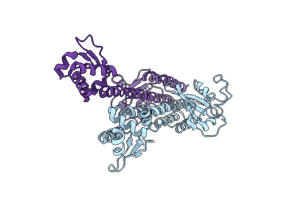
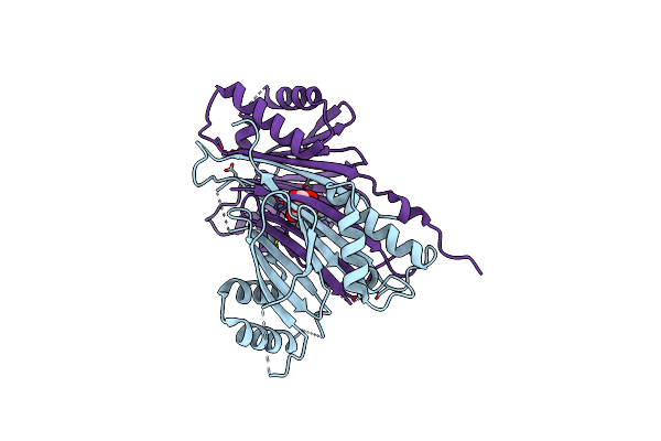
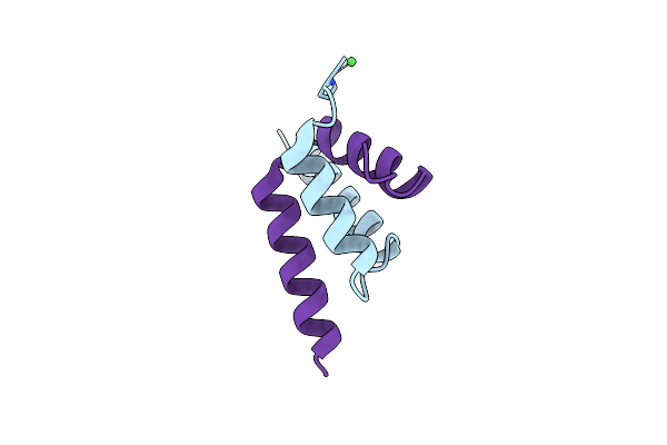
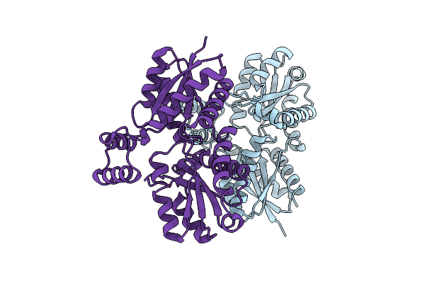
Structure Of The Phosphatase Domain Of The Cell Fate Determinant Spoiie From Bacillus Subtilis In A Crystal Form Without Domain Swapping
Organism: Bacillus subtilis
Method: X-RAY DIFFRACTION Resolution:2.45 Å Release Date: 2017-05-31 Classification: TRANSFERASE |
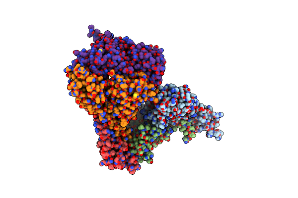 |
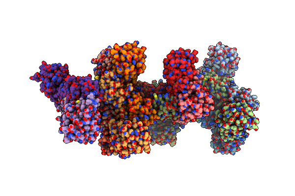
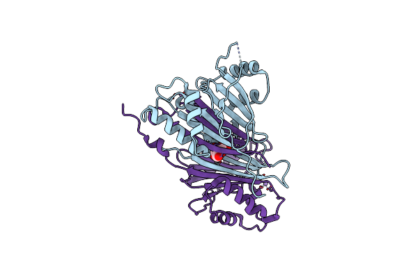
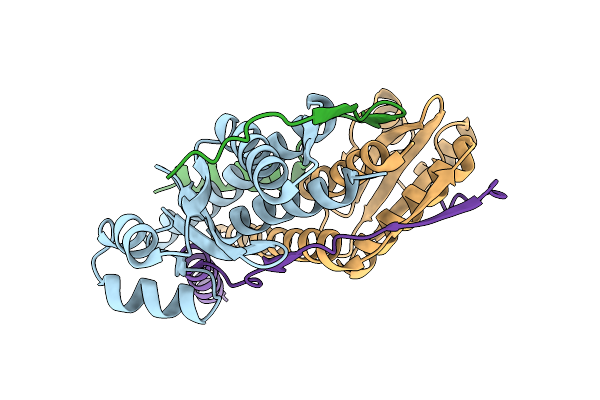
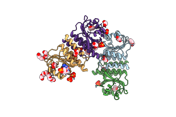
Structure Of The Pp2C Phosphatase Domain And A Fragment Of The Regulatory Domain Of The Cell Fate Determinant Spoiie From Bacillus Subtilis
Organism: Bacillus subtilis (strain 168)
Method: X-RAY DIFFRACTION Resolution:3.91 Å Release Date: 2017-05-31 Classification: HYDROLASE |
 |
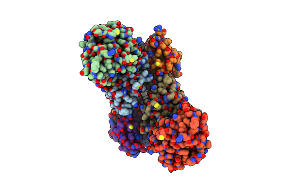
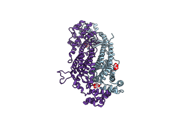
Organism: Bacillus subtilis (strain 168)
Method: X-RAY DIFFRACTION Resolution:3.00 Å Release Date: 2017-01-11 Classification: TRANSCRIPTION Ligands: SO4 |
 |
Organism: Bacillus subtilis (strain 168)
Method: X-RAY DIFFRACTION Resolution:3.00 Å Release Date: 2017-01-11 Classification: TRANSCRIPTION Ligands: ILE |
 |
Organism: Bacillus subtilis (strain 168)
Method: X-RAY DIFFRACTION Resolution:3.71 Å Release Date: 2017-01-11 Classification: TRANSCRIPTION |
 |
Organism: Bacillus subtilis
Method: X-RAY DIFFRACTION Resolution:4.50 Å Release Date: 2017-01-11 Classification: TRANSCRIPTION |
 |
Organism: Bacteriophage g20c
Method: X-RAY DIFFRACTION Resolution:1.98 Å Release Date: 2015-05-27 Classification: TRANSPORT PROTEIN Ligands: MPD |
 |
Organism: Bacillus subtilis
Method: X-RAY DIFFRACTION Resolution:2.26 Å Release Date: 2012-03-14 Classification: SIGNALING PROTEIN |
 |
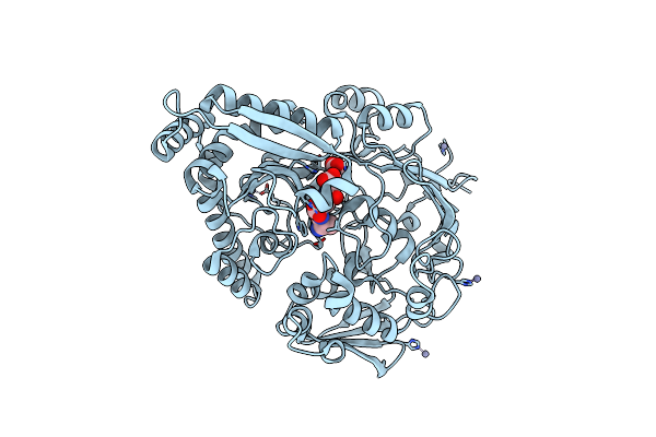
Structure Of The Phosphatase Domain Of The Cell Fate Determinant Spoiie From Bacillus Subtilis
Organism: Bacillus subtilis
Method: X-RAY DIFFRACTION Resolution:2.64 Å Release Date: 2011-12-07 Classification: HYDROLASE Ligands: MN, GL0, MAN |
 |
Structure Of The Phosphatase Domain Of The Cell Fate Determinant Spoiie From Bacillus Subtilis (Mn Presoaked)
Organism: Bacillus subtilis
Method: X-RAY DIFFRACTION Resolution:2.76 Å Release Date: 2011-12-07 Classification: HYDROLASE Ligands: MN, GL0 |
 |
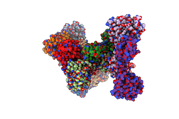
The Structure Of The Escherichia Coli Murein Tripeptide Binding Protein Mppa
Organism: Escherichia coli
Method: X-RAY DIFFRACTION Resolution:2.07 Å Release Date: 2011-07-06 Classification: PEPTIDE BINDING PROTEIN/PEPTIDE Ligands: MHI, ZN |
 |
Organism: Bacillus subtilis
Method: X-RAY DIFFRACTION Resolution:1.90 Å Release Date: 2011-06-15 Classification: TRANSCRIPTION |
 |
Organism: Bacillus subtilis
Method: X-RAY DIFFRACTION Resolution:2.27 Å Release Date: 2011-06-08 Classification: TRANSCRIPTION Ligands: NI |
 |
Organism: Bacillus subtilis
Method: X-RAY DIFFRACTION Resolution:2.50 Å Release Date: 2010-12-08 Classification: TOXIN/ANTITOXIN |
 |
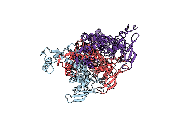
Structure Of The Proteolytic Domain Of The Human Mitochondrial Lon Protease
Organism: Homo sapiens
Method: X-RAY DIFFRACTION Resolution:2.00 Å Release Date: 2010-05-19 Classification: HYDROLASE |
 |
Crystal Structure Of Rph, An Exoribonuclease From Bacillus Anthracis At 1.7 A Resolution
Organism: Bacillus anthracis
Method: X-RAY DIFFRACTION Resolution:1.70 Å Release Date: 2009-02-10 Classification: TRANSFERASE Ligands: SO4 |
 |
Crystal Structure Of Ferrochelatase Hemh-1 From Bacillus Anthracis, Str. Ames
Organism: Bacillus anthracis
Method: X-RAY DIFFRACTION Resolution:2.10 Å Release Date: 2007-05-01 Classification: LYASE |
 |
Organism: Bacillus subtilis
Method: X-RAY DIFFRACTION Resolution:1.74 Å Release Date: 2007-04-17 Classification: TRANSCRIPTION Ligands: SO4, PGE, GOL, PG4, P6G, PCG |
 |
Organism: Bacillus anthracis
Method: X-RAY DIFFRACTION Resolution:2.00 Å Release Date: 2007-04-17 Classification: LYASE Ligands: MLI |