
Search Count: 25
 |
Organism: Homo sapiens
Method: X-RAY DIFFRACTION Resolution:1.30 Å Release Date: 2023-05-17 Classification: DNA BINDING PROTEIN Ligands: ZN |
 |
Organism: Homo sapiens
Method: X-RAY DIFFRACTION Resolution:2.50 Å Release Date: 2023-05-17 Classification: DNA BINDING PROTEIN Ligands: ZN |
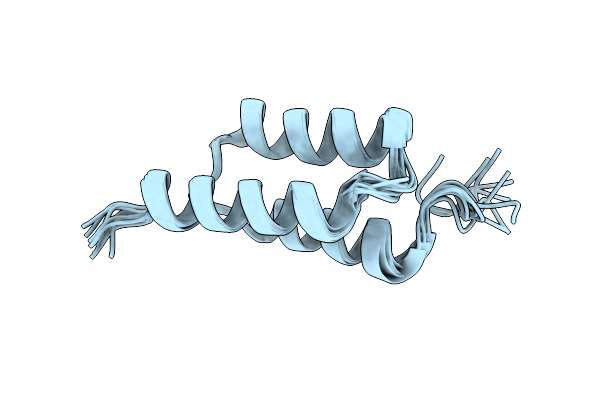 |
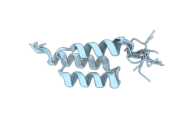
Backbone-Modified Variant Of The B Domain Of Staphylococcal Protein A: Beta3- And Acpc-Residues In Helix 2
Organism: Staphylococcus aureus
Method: SOLUTION NMR Release Date: 2023-05-03 Classification: DE NOVO PROTEIN |
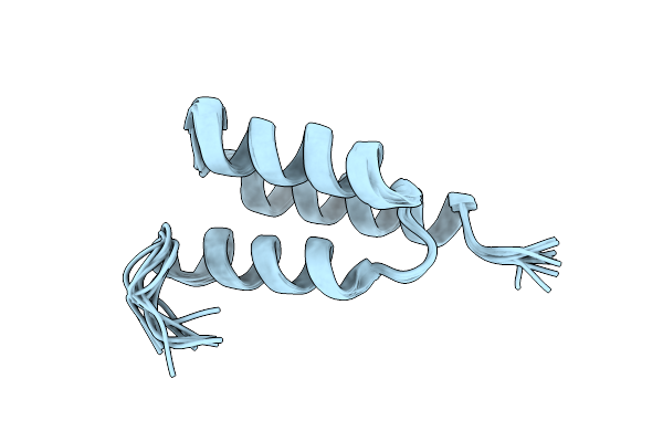 |
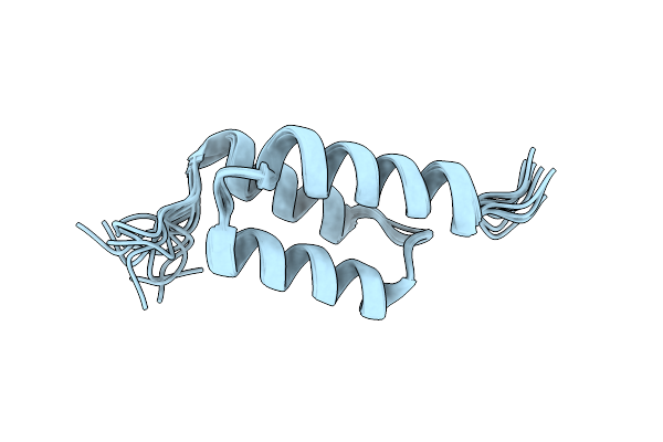
Backbone-Modified Variant Of The B Domain Of Staphylococcal Protein A: Beta3-Residues In Helix 2
Organism: Staphylococcus aureus
Method: SOLUTION NMR Release Date: 2022-12-21 Classification: DE NOVO PROTEIN |
 |
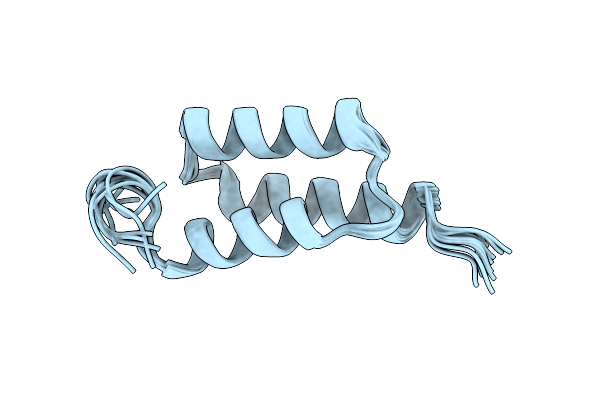
Backbone-Modified Variant Of The B Domain Of Staphylococcal Protein A: Aib Residues In Helix 2
Organism: Staphylococcus aureus
Method: SOLUTION NMR Release Date: 2022-12-21 Classification: DE NOVO PROTEIN |
 |
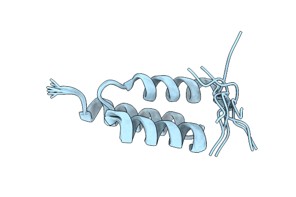
Backbone-Modified Variant Of The B Domain Of Staphylococcal Protein A: Beta3-Residues In Helix 3
Organism: Staphylococcus aureus
Method: SOLUTION NMR Release Date: 2022-12-21 Classification: DE NOVO PROTEIN |
 |
Backbone-Modified Variant Of The B Domain Of Staphylococcal Protein A: Aib Residues In Helix 3
Organism: Staphylococcus aureus
Method: SOLUTION NMR Release Date: 2022-12-21 Classification: DE NOVO PROTEIN |
 |
Organism: Staphylococcus aureus
Method: SOLUTION NMR Release Date: 2022-11-23 Classification: DE NOVO PROTEIN |
 |
Organism: Homo sapiens
Method: X-RAY DIFFRACTION Resolution:3.25 Å Release Date: 2019-11-20 Classification: TRANSFERASE Ligands: L1H |
 |
Organism: Homo sapiens
Method: X-RAY DIFFRACTION Resolution:2.73 Å Release Date: 2019-11-20 Classification: TRANSFERASE Ligands: L1K, DMS |
 |
Organism: Homo sapiens
Method: X-RAY DIFFRACTION Resolution:2.67 Å Release Date: 2019-11-20 Classification: TRANSFERASE Ligands: L3Z, DMS |
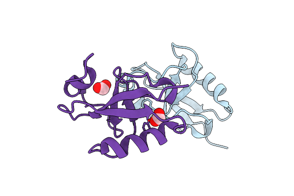 |
Crystal Structure Of The C-Terminal Domain Of Doublecortin (Tgdcx) From Toxoplasma Gondii Me49
Organism: Toxoplasma gondii
Method: X-RAY DIFFRACTION Resolution:2.00 Å Release Date: 2017-10-18 Classification: STRUCTURAL PROTEIN Ligands: FMT |
 |
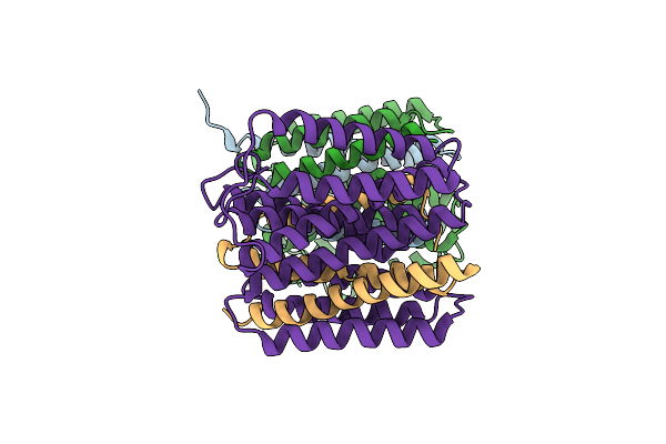
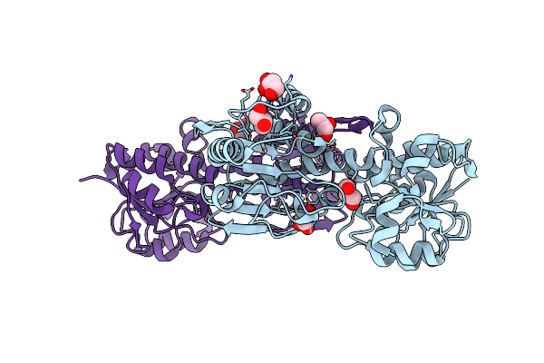
Critical Role Of Water Molecules For Proton Translocation Of The Membrane-Bound Transhydrogenase
Organism: Thermus thermophilus
Method: X-RAY DIFFRACTION Resolution:2.20 Å Release Date: 2017-05-10 Classification: OXIDOREDUCTASE Ligands: 1PE, PEG, OLC, BEN |
 |
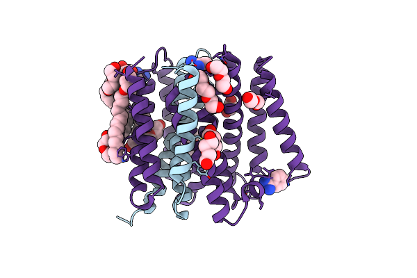
The Structure Of The Eukaryotic Purine/H+ Symporter, Uapa, In Complex With Xanthine
Organism: Aspergillus nidulans fgsc a4
Method: X-RAY DIFFRACTION Resolution:3.70 Å Release Date: 2016-04-27 Classification: TRANSPORT PROTEIN Ligands: XAN, LMT |
 |
Mechanism Of Transhydrogenase Coupling Proton Translocation And Hydride Transfer
Organism: Thermus thermophilus
Method: X-RAY DIFFRACTION Resolution:6.93 Å Release Date: 2015-01-28 Classification: MEMBRANE PROTEIN Ligands: NAP, NAD |
 |
Organism: Thermus thermophilus
Method: X-RAY DIFFRACTION Resolution:2.77 Å Release Date: 2014-12-31 Classification: MEMBRANE PROTEIN Ligands: HG |
 |
Crystal Structure Of Thermus Thermophilis Transhydrogeanse Domain Ii Dimer Semet Derivative
Organism: Thermus thermophilus
Method: X-RAY DIFFRACTION Resolution:2.89 Å Release Date: 2014-06-11 Classification: MEMBRANE PROTEIN |
 |
Mechanism Of Transhydrogenase Coupling Proton Translocation And Hydride Transfer
Organism: Thermus thermophilus
Method: X-RAY DIFFRACTION Resolution:3.08 Å Release Date: 2014-06-11 Classification: MEMBRANE PROTEIN |
 |
Crystal Structure Of The Alpha1 Dimer Of Thermus Thermophilus Transhydrogenase In P4(3)
Organism: Thermus thermophilus
Method: X-RAY DIFFRACTION Resolution:2.40 Å Release Date: 2014-02-05 Classification: OXIDOREDUCTASE Ligands: GOL |
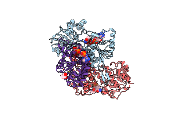 |
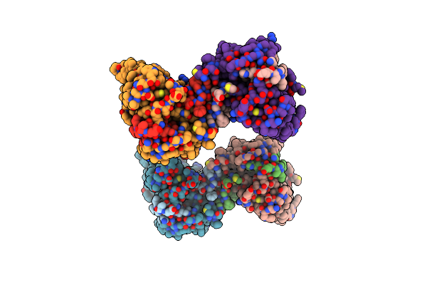
Crystal Structure Of Thermus Thermophilus Transhydrogenase Heterotrimeric Complex Of The Alpha1 Subunit Dimer With The Nadp Binding Domain (Domain Iii) Of The Beta Subunit
Organism: Thermus thermophilus
Method: X-RAY DIFFRACTION Resolution:2.41 Å Release Date: 2014-02-05 Classification: OXIDOREDUCTASE Ligands: NAD, CL, PGE, GOL, NAP |