
Search Count: 422
 |
Organism: Leptospira biflexa serovar patoc strain 'patoc 1 (paris)'
Method: X-RAY DIFFRACTION Resolution:2.20 Å Release Date: 2021-03-31 Classification: SIGNALING PROTEIN Ligands: GH3, MG, SO4, EDO |
 |
Organism: Leptospira biflexa serovar patoc (strain patoc 1 / atcc 23582 / paris)
Method: X-RAY DIFFRACTION Resolution:2.80 Å Release Date: 2021-03-31 Classification: SIGNALING PROTEIN Ligands: MG, GH3 |
 |
Organism: Leptospira biflexa serovar patoc strain 'patoc 1 (paris)'
Method: X-RAY DIFFRACTION Resolution:3.30 Å Release Date: 2021-03-31 Classification: SIGNALING PROTEIN Ligands: C2E, MG |
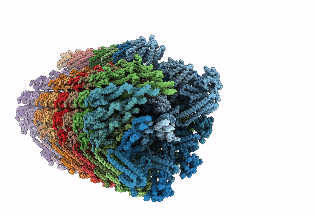 |
Rigid Body Fitting Of Flagellin Flab, And Flagellar Coiling Proteins, Fcpa And Fcpb, Into A 10 Angstrom Structure Of The Asymmetric Flagellar Filament Purified From Leptospira Biflexa Patoc Wt Cells Resolved Via Subtomogram Averaging
Organism: Leptospira biflexa serovar patoc (strain patoc 1 / atcc 23582 / paris)
Method: ELECTRON MICROSCOPY Release Date: 2020-03-25 Classification: STRUCTURAL PROTEIN |
 |
Organism: Leptospira biflexa serovar patoc (strain patoc 1 / atcc 23582 / paris)
Method: X-RAY DIFFRACTION Resolution:1.90 Å Release Date: 2020-01-29 Classification: PROTEIN FIBRIL Ligands: GOL, PO4, EPE |
 |
Organism: Leptospira biflexa serovar patoc (strain patoc 1 / atcc 23582 / paris)
Method: X-RAY DIFFRACTION Resolution:2.95 Å Release Date: 2020-01-29 Classification: PROTEIN FIBRIL Ligands: GOL |
 |
Flagellar Protein Fcpa From Leptospira Biflexa / Ab-Centered Monoclinic Form
Organism: Leptospira biflexa serovar patoc (strain patoc 1 / atcc 23582 / paris)
Method: X-RAY DIFFRACTION Resolution:2.50 Å Release Date: 2020-01-29 Classification: PROTEIN FIBRIL Ligands: GOL, IOD, PEG, TRS |
 |
Organism: Leptospira biflexa serovar patoc (strain patoc 1 / atcc 23582 / paris)
Method: X-RAY DIFFRACTION Resolution:2.78 Å Release Date: 2017-08-02 Classification: TRANSPORT PROTEIN |
 |
Organism: Leptospira biflexa serovar patoc (strain patoc 1 / atcc 23582 / paris)
Method: X-RAY DIFFRACTION Resolution:2.80 Å Release Date: 2017-08-02 Classification: TRANSPORT PROTEIN Ligands: MVC, BGC |
 |
Organism: Leptospira biflexa serovar patoc
Method: X-RAY DIFFRACTION Resolution:2.39 Å Release Date: 2014-09-10 Classification: MEMBRANE PROTEIN Ligands: OLC, MYS |
 |
Organism: Leptospira biflexa serovar patoc
Method: X-RAY DIFFRACTION Resolution:1.44 Å Release Date: 2014-05-14 Classification: SIGNALING PROTEIN Ligands: GOL, SO4 |
 |
NA
Organism: Leptospira biflexa serovar Patoc (strain Patoc 1 / Ames)
Method: Alphafold Release Date: Classification: NA Ligands: NA |
 |
NA
Organism: Leptospira biflexa serovar Patoc (strain Patoc 1 / Ames)
Method: Alphafold Release Date: Classification: NA Ligands: NA |
 |
NA
Organism: Leptospira biflexa serovar Patoc (strain Patoc 1 / Ames)
Method: Alphafold Release Date: Classification: NA Ligands: NA |
 |
NA
Organism: Leptospira biflexa serovar Patoc (strain Patoc 1 / Ames)
Method: Alphafold Release Date: Classification: NA Ligands: NA |
 |
NA
Organism: Leptospira biflexa serovar Patoc (strain Patoc 1 / Ames)
Method: Alphafold Release Date: Classification: NA Ligands: NA |
 |
NA
Organism: Leptospira biflexa serovar Patoc (strain Patoc 1 / Ames)
Method: Alphafold Release Date: Classification: NA Ligands: NA |
 |
NA
Organism: Leptospira biflexa serovar Patoc (strain Patoc 1 / Ames)
Method: Alphafold Release Date: Classification: NA Ligands: NA |
 |
NA
Organism: Leptospira biflexa serovar Patoc (strain Patoc 1 / Ames)
Method: Alphafold Release Date: Classification: NA Ligands: NA |
 |
NA
Organism: Leptospira biflexa serovar Patoc (strain Patoc 1 / Ames)
Method: Alphafold Release Date: Classification: NA Ligands: NA |