
Search Count: 13
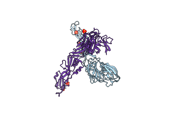 |
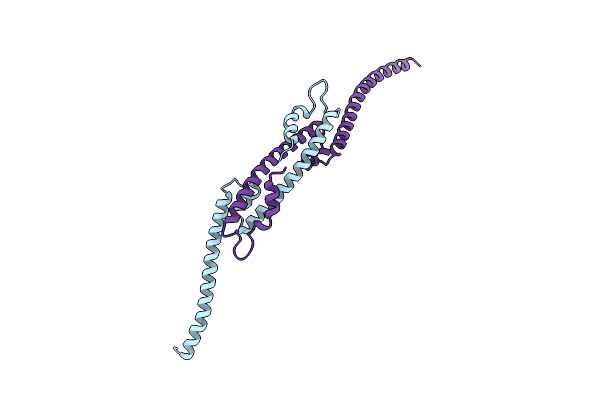
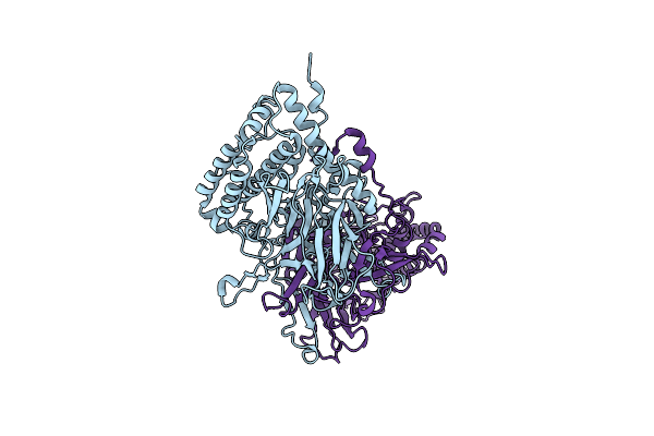
Structure Of The Yellow Fever Virus (Asibi Strain) Dimeric Envelope Protein
Organism: Yellow fever virus
Method: X-RAY DIFFRACTION Resolution:3.48 Å Release Date: 2023-08-09 Classification: VIRAL PROTEIN Ligands: SO4 |
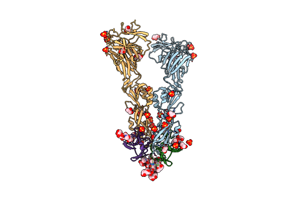 |
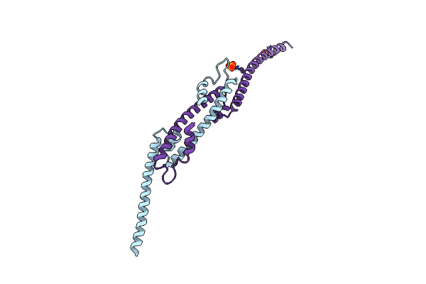
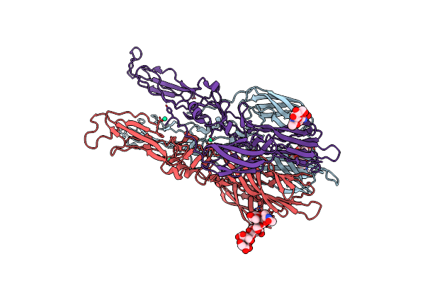
Crystal Structure Of The Precursor Membrane Protein-Envelope Protein Heterodimer From The Yellow Fever Virus
Organism: Yellow fever virus
Method: X-RAY DIFFRACTION Resolution:2.70 Å Release Date: 2018-10-31 Classification: VIRAL PROTEIN Ligands: SO4, GOL |
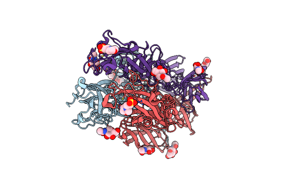 |
Structure Of Rvfv Envelope Protein Gc In Postfusion Conformation In Complex With Mes
Organism: Rift valley fever virus
Method: X-RAY DIFFRACTION Resolution:2.50 Å Release Date: 2017-11-01 Classification: VIRAL PROTEIN Ligands: NAG, MES |
 |
Structure Of Rvfv Envelope Protein Gc In Postfusion Conformation In Complex With 1,2-Dipropionyl-Sn-Glycero-3-Phosphocholine
Organism: Rift valley fever virus
Method: X-RAY DIFFRACTION Resolution:2.30 Å Release Date: 2017-11-01 Classification: VIRAL PROTEIN Ligands: NAG, 43Y |
 |
Structural Intermediates In The Fusion Associated Transition Of Vesiculovirus Glycoprotein
Organism: Chandipura virus
Method: X-RAY DIFFRACTION Resolution:3.00 Å Release Date: 2016-12-14 Classification: VIRAL PROTEIN Ligands: SO4, NAG |
 |
Crystal Structure Of Fragment 1600-1733 Of Hsv1 Ul36 In The Presence Of 1M Potassium Iodide
Organism: Human herpesvirus 1(type 1 / strain 17)
Method: X-RAY DIFFRACTION Resolution:2.60 Å Release Date: 2015-02-18 Classification: HYDROLASE Ligands: IOD |
 |
Organism: Herpes simplex virus (type 1 / strain 17)
Method: X-RAY DIFFRACTION Resolution:2.75 Å Release Date: 2015-02-18 Classification: HYDROLASE Ligands: PO4 |
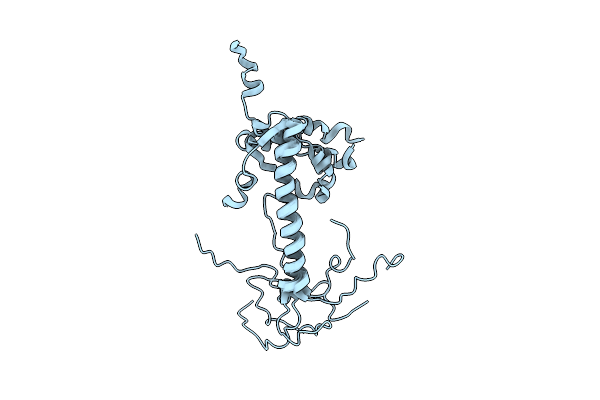 |
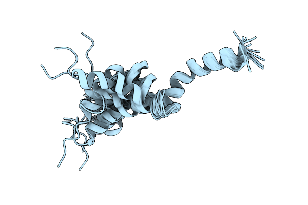
Nmr Structure Of The C-Terminal Domain Of Vp7 In Membrane Mimicking Micelles
|
 |
Organism: Human rotavirus a
Method: SOLUTION NMR Release Date: 2011-03-30 Classification: VIRAL PROTEIN |
 |
Organism: Rabbit picobirnavirus
Method: X-RAY DIFFRACTION Resolution:3.40 Å Release Date: 2008-12-16 Classification: VIRUS |
 |
Nmr Structure Of Pep46 From The Infectious Bursal Disease Virus (Ibdv) In Dodecylphosphocholin (Dpc).
|
 |
Crystal Structure Of The Homotrimer Of Fusion Glycoprotein E1 From Semliki Forest Virus.
Organism: Semliki forest virus
Method: X-RAY DIFFRACTION Resolution:3.20 Å Release Date: 2004-01-27 Classification: VIRAL PROTEIN Ligands: BR, HO, PO4 |
 |
Organism: Bovine rotavirus
Method: X-RAY DIFFRACTION Resolution:1.95 Å Release Date: 2001-04-13 Classification: VIRAL PROTEIN Ligands: ZN, CL, CA |