
Search Count: 82
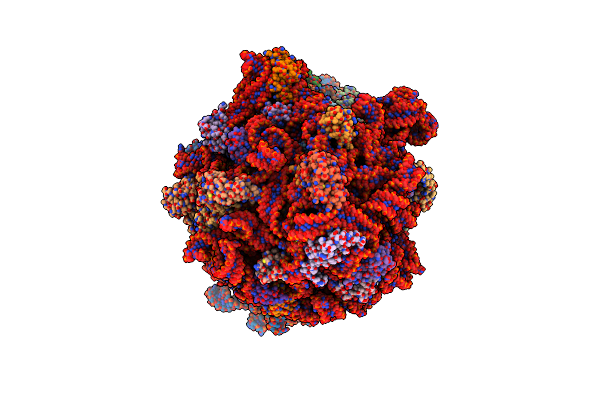 |
Principles Of Ion Binding To Rna Inferred From The Analysis Of A 1.55 Angstrom Resolution Bacterial Ribosome Structure - Part I: Mg2+
Organism: Escherichia coli b
Method: ELECTRON MICROSCOPY Release Date: 2025-05-14 Classification: RIBOSOME Ligands: K, MG, ZN |
 |
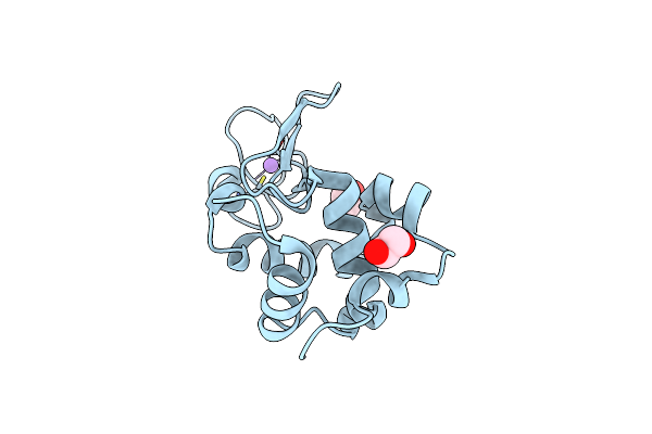
Dark Structure Of The Human Adenosine A2A Receptor Bound To Synthetic Photoswitch "Stilswitch3" Determined By Serial Synchrotron Crystallography
Organism: Homo sapiens
Method: X-RAY DIFFRACTION Resolution:2.65 Å Release Date: 2025-01-08 Classification: MEMBRANE PROTEIN Ligands: OLA, A1H3H, CLR, NA |
 |
Crystal Structure Of The Adenosine A2A Receptor In Complex With Istradefylline
Organism: Homo sapiens
Method: X-RAY DIFFRACTION Resolution:1.94 Å Release Date: 2025-01-08 Classification: MEMBRANE PROTEIN Ligands: JQ9, CLR, OLA, NA |
 |
Crystal Structure Of The Adenosine A2A Receptor In Complex With The Synthetic Photoswitch 'Azoswitch2
Organism: Homo sapiens
Method: X-RAY DIFFRACTION Resolution:2.20 Å Release Date: 2025-01-08 Classification: MEMBRANE PROTEIN Ligands: A1H3L, CLR, OLA, NA |
 |
Crystal Structure Of The Adenosine A2A Receptor In Complex With The Synthetic Photoswitch 'Stilswitch1
Organism: Homo sapiens
Method: X-RAY DIFFRACTION Resolution:2.25 Å Release Date: 2025-01-08 Classification: MEMBRANE PROTEIN Ligands: A1H3J, CLR, OLA, OLC, OLB, NA |
 |
Crystal Structure Of The Adenosine A2A Receptor In Complex With The Synthetic Photoswitch 'Stilswitch2
Organism: Homo sapiens
Method: X-RAY DIFFRACTION Resolution:2.31 Å Release Date: 2025-01-08 Classification: MEMBRANE PROTEIN Ligands: A1H3I, CLR, OLC, OLA, NA |
 |
Crystal Structure Of The Adenosine A2A Receptor In Complex With The Synthetic Photoswitch 'Stilswitch3
Organism: Homo sapiens
Method: X-RAY DIFFRACTION Resolution:2.05 Å Release Date: 2025-01-08 Classification: MEMBRANE PROTEIN Ligands: A1H3H, CLR, OLA, NA |
 |
Crystal Structure Of The Adenosine A2A Receptor In Complex With The Synthetic Photoswitch 'Stilswitch4
Organism: Homo sapiens
Method: X-RAY DIFFRACTION Resolution:2.20 Å Release Date: 2025-01-08 Classification: MEMBRANE PROTEIN Ligands: A1H3K, CLR, OLA, OLC, NA |
 |
Dark Structure Of The Human Adenosine A2A Receptor Bound To Synthetic Photoswitch 'Stilswitch2' Determined By Serial Synchrotron Crystallography
Organism: Homo sapiens
Method: X-RAY DIFFRACTION Resolution:2.45 Å Release Date: 2025-01-08 Classification: MEMBRANE PROTEIN Ligands: A1H3I, CLR, OLA, OLB, OLC, NA |
 |
Steady State Structure Of The Human Adenosine A2A Receptor Bound To Synthetic Photoswitch 'Stilswitch2' Determined By Serial Synchrotron Crystallography
Organism: Homo sapiens
Method: X-RAY DIFFRACTION Resolution:2.80 Å Release Date: 2025-01-08 Classification: MEMBRANE PROTEIN Ligands: A1H3I, CLR, OLA, NA |
 |
Steady State Structure Of The Human Adenosine A2A Receptor Bound To Synthetic Photoswitch 'Stilswitch3' Determined By Serial Synchrotron Crystallography
Organism: Homo sapiens, Escherichia coli
Method: X-RAY DIFFRACTION Resolution:3.05 Å Release Date: 2025-01-08 Classification: MEMBRANE PROTEIN Ligands: A1H3H, OLA, NA |
 |
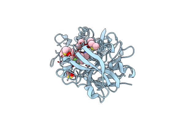
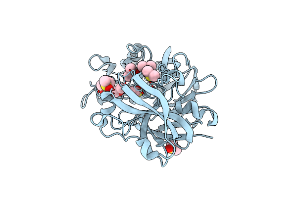
Lysozyme Structure Solved From Serial Crystallography Data Collected At 2 Khz For 5 Seconds With Jungfrau Detector At Maxiv
Organism: Gallus gallus
Method: X-RAY DIFFRACTION Resolution:2.05 Å Release Date: 2023-10-18 Classification: HYDROLASE Ligands: EDO, CL |
 |
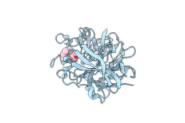
Lysozyme Structure Solved From Serial Crystallography Data Collected At 2 Khz With Jungfrau Detector At Maxiv
Organism: Gallus gallus
Method: X-RAY DIFFRACTION Resolution:1.70 Å Release Date: 2023-10-18 Classification: HYDROLASE Ligands: EDO, CL |
 |
Lysozyme Structure Solved From Serial Crystallography Data Collected At 1 Khz With Jungfrau Detector At Maxiv
Organism: Gallus gallus
Method: X-RAY DIFFRACTION Resolution:1.58 Å Release Date: 2023-10-18 Classification: HYDROLASE Ligands: EDO, CL, NA |
 |
Lysozyme Structure Solved From Serial Crystallography Data Collected At 100 Hz With Jungfrau Detector At Maxiv
Organism: Gallus gallus
Method: X-RAY DIFFRACTION Resolution:1.60 Å Release Date: 2023-10-18 Classification: HYDROLASE Ligands: EDO, CL, NA |
 |
Organism: Cryphonectria parasitica
Method: X-RAY DIFFRACTION Resolution:1.39 Å Release Date: 2022-08-17 Classification: HYDROLASE Ligands: PG5, DMS, U1H |
 |
Organism: Cryphonectria parasitica
Method: X-RAY DIFFRACTION Resolution:1.41 Å Release Date: 2022-08-17 Classification: HYDROLASE Ligands: PG5 |
 |
Organism: Cryphonectria parasitica
Method: X-RAY DIFFRACTION Resolution:1.41 Å Release Date: 2022-08-17 Classification: HYDROLASE Ligands: DMS, PG5 |
 |
Organism: Cryphonectria parasitica
Method: X-RAY DIFFRACTION Resolution:1.41 Å Release Date: 2022-08-17 Classification: HYDROLASE Ligands: DMS, PG5 |
 |
Organism: Cryphonectria parasitica
Method: X-RAY DIFFRACTION Resolution:1.39 Å Release Date: 2022-08-17 Classification: HYDROLASE Ligands: DMS, PG5 |