
Search Count: 28
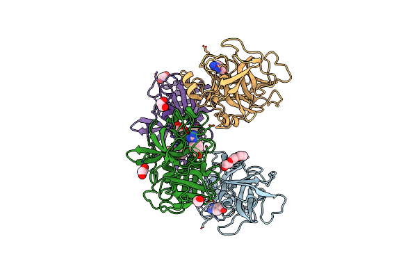 |
Organism: Homo sapiens
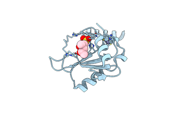
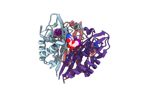
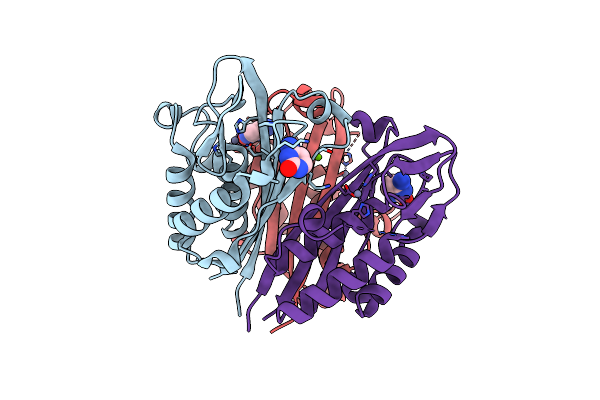
Method: X-RAY DIFFRACTION Resolution:2.19 Å Release Date: 2020-10-21 Classification: HYDROLASE Ligands: GBS, PGE, EDO, GOL |
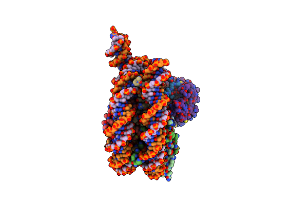 |
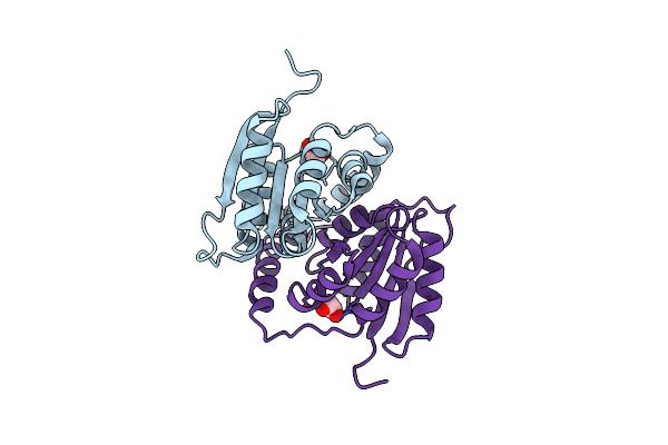
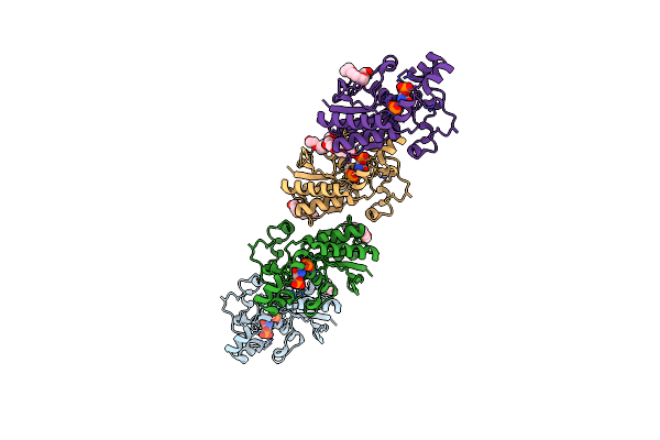
Cryo-Em Structure Of Singly-Bound Snf2H-Nucleosome Complex With Snf2H Bound At Shl-2
Organism: Xenopus laevis, Homo sapiens
Method: ELECTRON MICROSCOPY Release Date: 2019-07-17 Classification: DNA BINDING PROTEIN/DNA Ligands: ADP |
 |
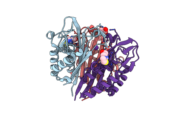
Crystal Structure Of Ribose-5-Phosphate Isomerase B Rpib From Coccidioides Immitis Semi-Covalently Bound To Malonic Acid
Organism: Coccidioides immitis
Method: X-RAY DIFFRACTION Resolution:1.70 Å Release Date: 2011-06-29 Classification: ISOMERASE/ISOMERASE INHIBITOR Ligands: MLA, EDO, CL |
 |
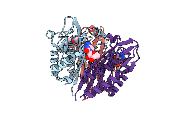
Crystal Structure Of A Ribose-5-Phosphate Isomerase B Rpib From Coccidioides Immitis Bound To Phosphate
Organism: Coccidioides immitis
Method: X-RAY DIFFRACTION Resolution:1.80 Å Release Date: 2011-06-22 Classification: ISOMERASE Ligands: PO4, EDO, CL |
 |
Crystal Structure Of A Zinc-Containing Hit Family Protein From Encephalitozoon Cuniculi
Organism: Encephalitozoon cuniculi
Method: X-RAY DIFFRACTION Resolution:1.85 Å Release Date: 2011-03-30 Classification: HYDROLASE Ligands: ZN, SO4, MPD |
 |
Crystal Structure Of A Putative Ribose-5-Phosphate Isomerase From Coccidioides Immitis Solved By Combined Iodide Ion Sad And Mr
Organism: Coccidioides immitis
Method: X-RAY DIFFRACTION Resolution:1.90 Å Release Date: 2011-02-09 Classification: ISOMERASE Ligands: EDO, IOD |
 |
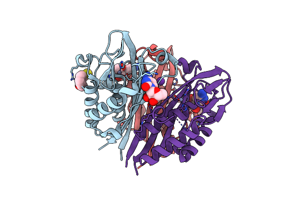
Crystal Structure Of 2-C-Methyl-D-Erythritol 2,4-Cyclodiphosphate Synthase From Burkholderia Pseudomallei Bound To Cytidine, Fol795 And Fol955
Organism: Burkholderia pseudomallei
Method: X-RAY DIFFRACTION Resolution:1.70 Å Release Date: 2011-02-09 Classification: LYASE Ligands: ZN, CL, CTN, MSR, 795, K |
 |
Crystal Structure Of 2-C-Methyl-D-Erythritol 2,4-Cyclodiphosphate Synthase From Burkholderia Pseudomallei With Cytidine And Fol694, 2-(Thiophen-2-Yl)Phenyl Methanol
Organism: Burkholderia pseudomallei
Method: X-RAY DIFFRACTION Resolution:1.70 Å Release Date: 2010-11-10 Classification: LYASE Ligands: CL, ZN, CTN, F69, K |
 |
Crystal Structure Of 2-C-Methyl-D-Erythritol 2,4-Cyclodiphosphate Synthase From Burkholderia Pseudomallei With Cytidine And Fol955, 4-(1H-Imidazol)-1-Yl)Phenol
Organism: Burkholderia pseudomallei
Method: X-RAY DIFFRACTION Resolution:1.95 Å Release Date: 2010-10-20 Classification: LYASE Ligands: CL, ZN, MSR, CTN, K |
 |
Crystal Structure Of 2C-Methyl-D-Erythritol 2,4-Cyclodiphosphate Synthase From Burkholderia Pseudomallei With Cytosine And Fol Fragment 717, Imidazo[2,1-B][1,3]Thiazol-6-Ylmethanol
Organism: Burkholderia pseudomallei
Method: X-RAY DIFFRACTION Resolution:1.80 Å Release Date: 2010-04-07 Classification: LYASE Ligands: ZN, CYT, 717, TRS |
 |
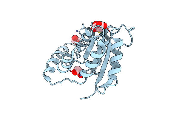
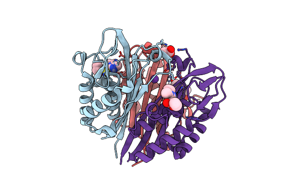
Crystal Structure Of Phosphoglyceromutase From Burkholderia Pseudomallei 1710B With Bound Malonic Acid
Organism: Burkholderia pseudomallei
Method: X-RAY DIFFRACTION Resolution:2.10 Å Release Date: 2010-02-09 Classification: ISOMERASE |
 |
Crystal Structure Of 2C-Methyl-D-Erythritol 2,4-Cyclodiphosphate Synthase From Burkholderia Pseudomallei In Complex With 5'-Iodo-Cytosine
Organism: Burkholderia pseudomallei
Method: X-RAY DIFFRACTION Resolution:1.85 Å Release Date: 2009-10-13 Classification: LYASE Ligands: ZN, I5A, CL, K |
 |
Co-Crystal Structure Of 2C-Methyl-D-Erythritol 2,4-Cyclodiphosphate Synthase From Burkholderia Pseudomallei With Fol Fragment 535, Ethyl 3-Methyl-5,6-Dihydroimidazo[2,1-B][1,3]Thiazole-2-Carboxylate
Organism: Burkholderia pseudomallei
Method: X-RAY DIFFRACTION Resolution:1.70 Å Release Date: 2009-10-06 Classification: LYASE Ligands: ZN, 535, CL, K, ACT |
 |
Crystal Structure Of 2C-Methyl-D-Erythritol-2,4-Cyclodiphosphate Synthase From Burkholderia Pseudomallei With Fol Fragment 8395
Organism: Burkholderia pseudomallei
Method: X-RAY DIFFRACTION Resolution:1.69 Å Release Date: 2009-09-29 Classification: LYASE Ligands: ZN, HHV, GOL |
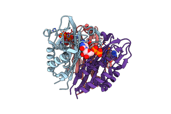 |
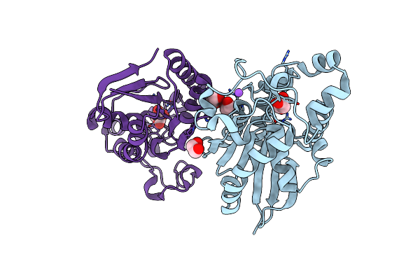
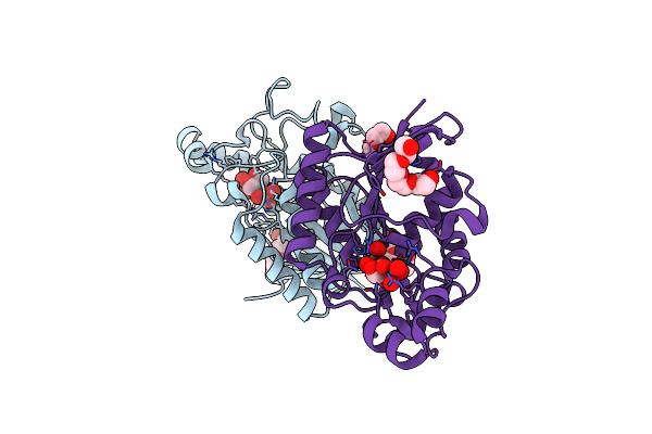
Crystal Structure Of 2C-Methyl-D-Erythritol 2,4-Cyclodiphosphate Synthase From Burkholderia Pseudomallei With Bound Ctp And Cdp
Organism: Burkholderia pseudomallei
Method: X-RAY DIFFRACTION Resolution:2.10 Å Release Date: 2009-08-18 Classification: LYASE Ligands: ZN, CDP, CTP, GOL |
 |
Crystal Structure Of 2C-Methyl-D-Erythritol 2,4-Cyclodiphosphate Synthase From Burkholderia Pseudomallei With Cytosine
Organism: Burkholderia pseudomallei
Method: X-RAY DIFFRACTION Resolution:2.30 Å Release Date: 2009-08-18 Classification: LYASE Ligands: ZN, CYT, CL, MG |
 |
Crystal Structure Of 2C-Methyl-D-Erythritol 2,4-Cyclodiphosphate Synthase From Burkholderia Pseudomallei With Fol Fragment 717, Imidazo[2,,1-B][1,3]Thiazol-6-Ylmethanol
Organism: Burkholderia pseudomallei
Method: X-RAY DIFFRACTION Resolution:2.07 Å Release Date: 2009-08-18 Classification: LYASE Ligands: ZN, 717, CL, ACT, K |
 |
Crystal Structure Of 2C-Methyl-D-Erythritol 2,4-Cyclodiphosphate Synthase From Burkholderia Pseudomallei With Cytidine
Organism: Burkholderia pseudomallei
Method: X-RAY DIFFRACTION Resolution:2.10 Å Release Date: 2009-08-04 Classification: LYASE Ligands: CTN, ZN, CL, MG |
 |
Crystal Structure Of Phosphoglyceromutase From Burkholderia Pseudomallei With Vanadate And Glycerol
Organism: Burkholderia pseudomallei
Method: X-RAY DIFFRACTION Resolution:1.93 Å Release Date: 2009-04-07 Classification: ISOMERASE Ligands: VO4, GOL, PG4 |
 |
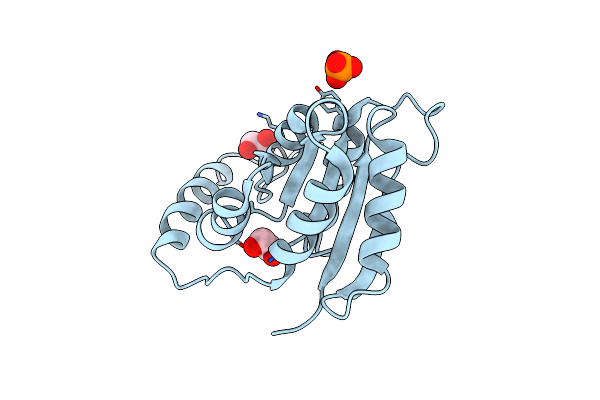
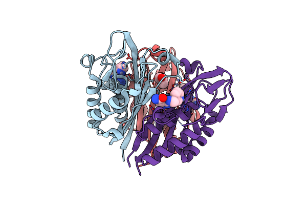
Crystal Structure Of Phosphoglyceromutase From Burkholderia Pseudomallei With 2-Phosphoserine
Organism: Burkholderia pseudomallei
Method: X-RAY DIFFRACTION Resolution:1.50 Å Release Date: 2009-03-31 Classification: ISOMERASE Ligands: PG4, PO3, SEP |