
Search Count: 98
 |
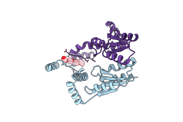
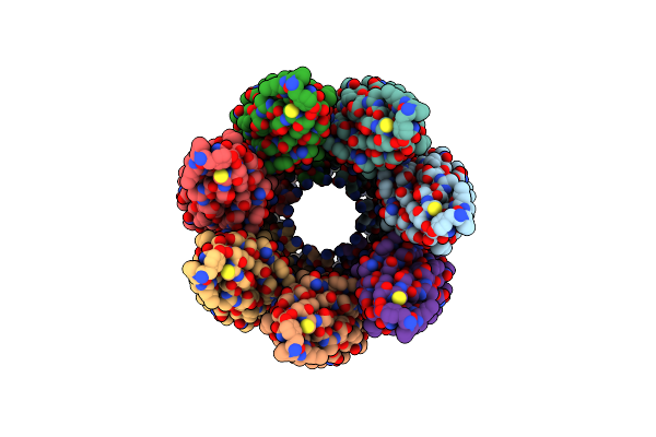
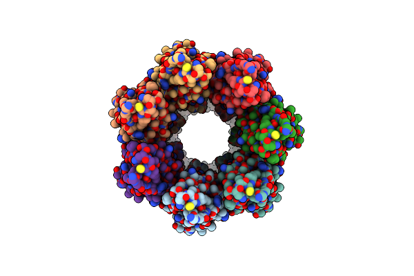
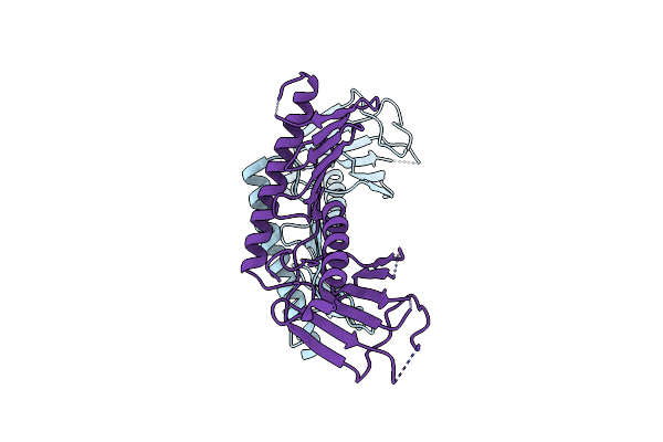
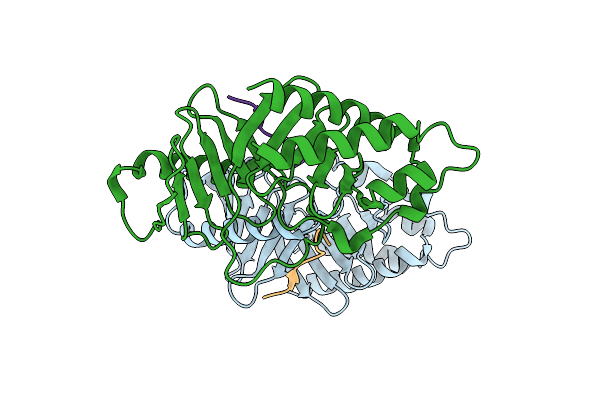
Crystal Structure Of N-Terminal Domain Of Hypothetical Protein Rv1421 From Mycobacterium Tuberculosis H37Rv In Complex With Uridine Diphosphate N-Acetyl Glucosamine
Organism: Mycobacterium tuberculosis h37rv
Method: X-RAY DIFFRACTION Release Date: 2025-09-03 Classification: HYDROLASE Ligands: UD1, P6G |
 |
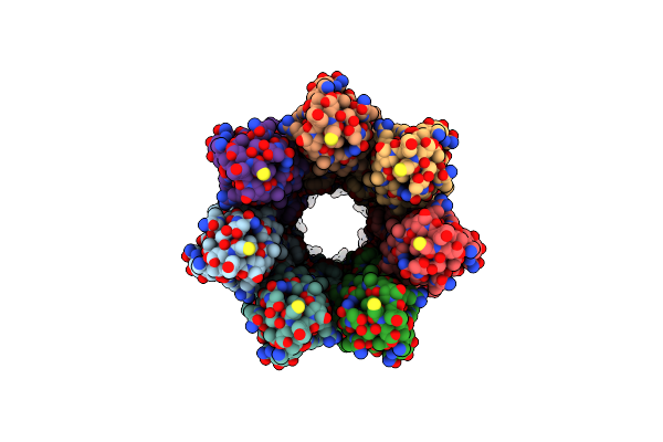
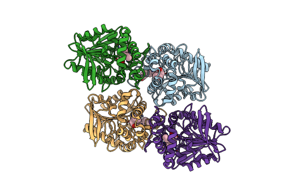
Crystal Structure Of N-Terminal Domain Of Hypothetical Protein Rv1421 From Mycobacterium Tuberculosis H37Rv
Organism: Mycobacterium tuberculosis h37rv
Method: X-RAY DIFFRACTION Release Date: 2025-04-30 Classification: HYDROLASE Ligands: P6G |
 |
Cryoem Structure Of The Neck (1403-2314) In The Grappling Hook Protein A (Ghpa) In The Bacterium Aureispira Sp. Ccb-Qb1
Organism: Aureispira sp. ccb-qb1
Method: ELECTRON MICROSCOPY Release Date: 2024-10-16 Classification: CELL ADHESION |
 |
Cryoem Structure Of The Fragment-3 (2061-2397) In The Grappling Hook Protein A (Ghpa) In The Bacterium Aureispira Sp. Ccb-Qb1
Organism: Aureispira sp. ccb-qb1
Method: ELECTRON MICROSCOPY Release Date: 2024-10-16 Classification: CELL ADHESION |
 |
Cryoem Structure Of The Fragment-1 (3402-3733) In The Grappling Hook Protein A (Ghpa) In The Bacterium Aureispira Sp. Ccb-Qb1
Organism: Aureispira sp. ccb-qb1
Method: ELECTRON MICROSCOPY Release Date: 2024-10-16 Classification: CELL ADHESION |
 |
Cryoem Structure Of The Fragment-5 (2393-2807) In The Grappling Hook Protein A (Ghpa) In The Bacterium Aureispira Sp. Ccb-Qb1
Organism: Aureispira sp. ccb-qb1
Method: ELECTRON MICROSCOPY Release Date: 2024-10-16 Classification: CELL ADHESION |
 |
Cryoem Structure Of The Fragment-6 (3571-4078) In The Grappling Hook Protein A (Ghpa) In The Bacterium Aureispira Sp. Ccb-Qb1
Organism: Aureispira sp. ccb-qb1
Method: ELECTRON MICROSCOPY Release Date: 2024-10-16 Classification: CELL ADHESION |
 |
Cryoem Structure Of The Fragment-4 (4074-4421) In The Grappling Hook Protein A (Ghpa) In The Bacterium Aureispira Sp. Ccb-Qb1
Organism: Aureispira sp. ccb-qb1
Method: ELECTRON MICROSCOPY Release Date: 2024-10-16 Classification: CELL ADHESION |
 |
Cryoem Structure Of The Base (4151-5009) In The Grappling Hook Protein A (Ghpa) In The Bacterium Aureispira Sp. Ccb-Qb1
Organism: Aureispira sp. ccb-qb1
Method: ELECTRON MICROSCOPY Release Date: 2024-10-16 Classification: CELL ADHESION |
 |
Cryoem Structure Of The Fragment-2 (2892-3236) In The Grappling Hook Protein A (Ghpa) In The Bacterium Aureispira Sp. Ccb-Qb1
Organism: Aureispira sp. ccb-qb1
Method: ELECTRON MICROSCOPY Release Date: 2024-10-16 Classification: CELL ADHESION |
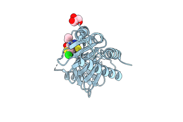 |
Organism: Homo sapiens
Method: X-RAY DIFFRACTION Resolution:1.65 Å Release Date: 2023-08-16 Classification: TRANSFERASE Ligands: GOL, VIH |
 |
Organism: Homo sapiens
Method: X-RAY DIFFRACTION Resolution:3.29 Å Release Date: 2023-07-19 Classification: STRUCTURAL PROTEIN |
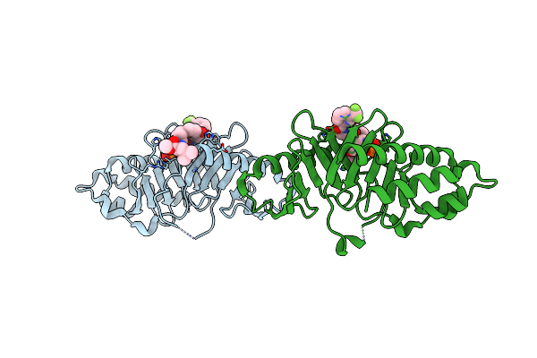 |
Crystal Structure Of Polo Box Domain In Complex With Cyclic Peptide Inhibitor
Organism: Homo sapiens, Synthetic construct
Method: X-RAY DIFFRACTION Resolution:1.85 Å Release Date: 2022-03-02 Classification: TRANSFERASE/Inhibitor |
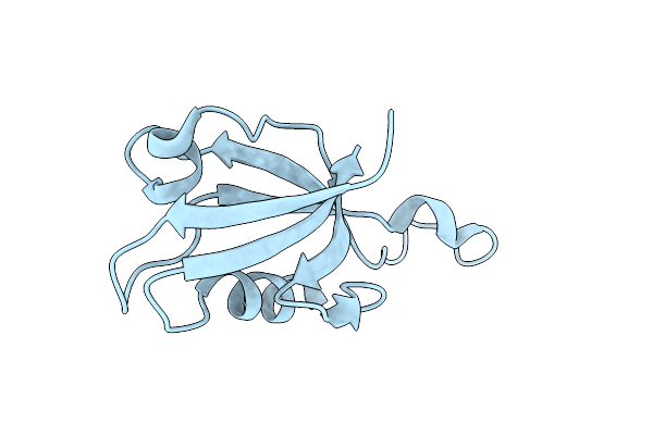 |
Organism: Homo sapiens
Method: X-RAY DIFFRACTION Resolution:1.15 Å Release Date: 2019-10-02 Classification: STRUCTURAL PROTEIN |
 |
Crystal Structure Of The Cryptic Polo Box Domain Of Human Activated Plk4 Variant 1
Organism: Homo sapiens
Method: X-RAY DIFFRACTION Resolution:2.64 Å Release Date: 2019-09-04 Classification: CELL CYCLE |
 |
Organism: Homo sapiens
Method: X-RAY DIFFRACTION Resolution:3.71 Å Release Date: 2019-09-04 Classification: CELL CYCLE |
 |
Organism: Homo sapiens
Method: X-RAY DIFFRACTION Resolution:2.50 Å Release Date: 2019-03-27 Classification: CELL CYCLE |
 |
Organism: Homo sapiens
Method: X-RAY DIFFRACTION Resolution:2.50 Å Release Date: 2019-03-27 Classification: CELL CYCLE |
 |
Crystal Structure Of Mouse Plk1-Pbd In Complex With Phosphopeptide From Hef1 (799-809)
Organism: Mus musculus, Homo sapiens
Method: X-RAY DIFFRACTION Resolution:2.90 Å Release Date: 2017-12-20 Classification: TRANSFERASE/PEPTIDE |
 |
Crystal Structure Of The E153Q Mutant Of The Cftr Inhibitory Factor Cif Containing The Adducted 16,17-Epdpe Hydrolysis Intermediate
Organism: Pseudomonas aeruginosa (strain ucbpp-pa14)
Method: X-RAY DIFFRACTION Resolution:1.80 Å Release Date: 2017-10-18 Classification: HYDROLASE Ligands: 7EZ |