
Search Count: 906
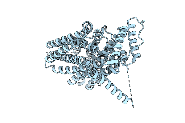 |
Organism: Homo sapiens
Method: ELECTRON MICROSCOPY Release Date: 2025-12-10 Classification: MEMBRANE PROTEIN |
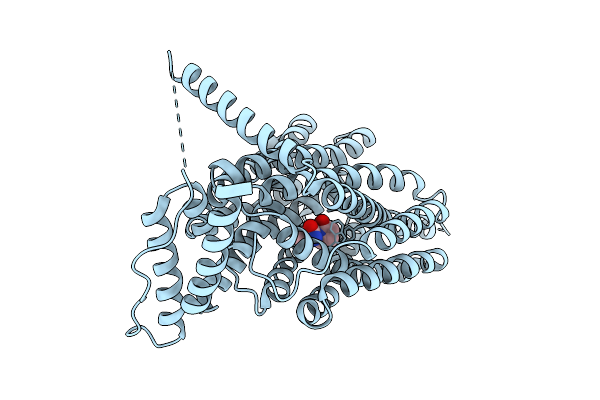 |
Organism: Homo sapiens
Method: ELECTRON MICROSCOPY Release Date: 2025-12-10 Classification: MEMBRANE PROTEIN Ligands: UKX |
 |
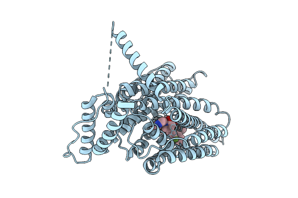
Organism: Homo sapiens
Method: ELECTRON MICROSCOPY Release Date: 2025-12-10 Classification: MEMBRANE PROTEIN Ligands: A1CCA |
 |
Organism: Homo sapiens
Method: ELECTRON MICROSCOPY Release Date: 2025-12-10 Classification: MEMBRANE PROTEIN Ligands: A1CCA, A1B1I |
 |
Organism: Homo sapiens
Method: ELECTRON MICROSCOPY Release Date: 2025-12-10 Classification: MEMBRANE PROTEIN Ligands: X3U |
 |
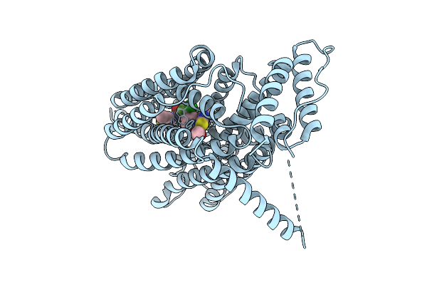
Cryo-Em Structure Of Human Sv2A In Complex With Levetiracetam And Ucb1244283
Organism: Homo sapiens
Method: ELECTRON MICROSCOPY Release Date: 2025-12-10 Classification: MEMBRANE PROTEIN Ligands: UKX, A1B1J |
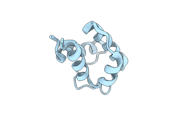 |
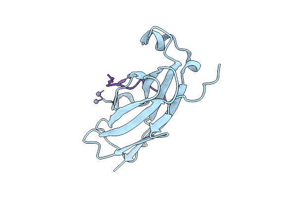
Solution Structure Of Holo Acyl Carrier Protein 2 (Apef) Of Aryl Polyene Biosynthesis From Acinetobacter Baumannii
Organism: Acinetobacter baumannii
Method: SOLUTION NMR Release Date: 2025-11-26 Classification: BIOSYNTHETIC PROTEIN |
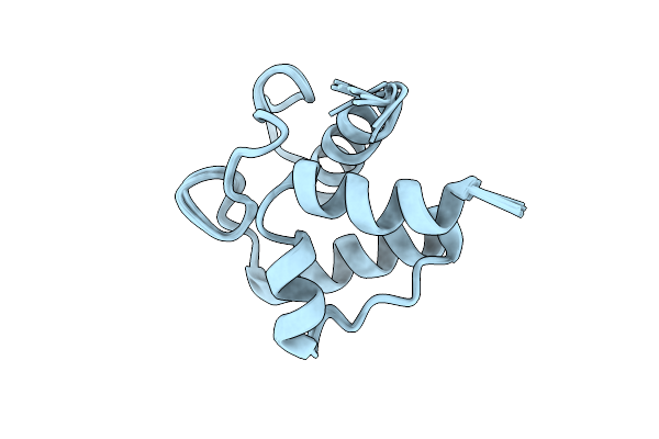 |
Solution Structure Of Holo Acyl Carrier Protein 1 (Apee) Of Aryl Polyene Biosynthesis From Acinetobacter Baumannii
Organism: Acinetobacter baumannii
Method: SOLUTION NMR Release Date: 2025-11-26 Classification: BIOSYNTHETIC PROTEIN |
 |
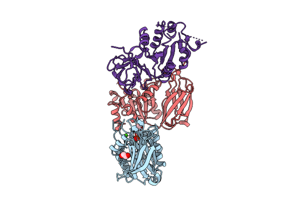
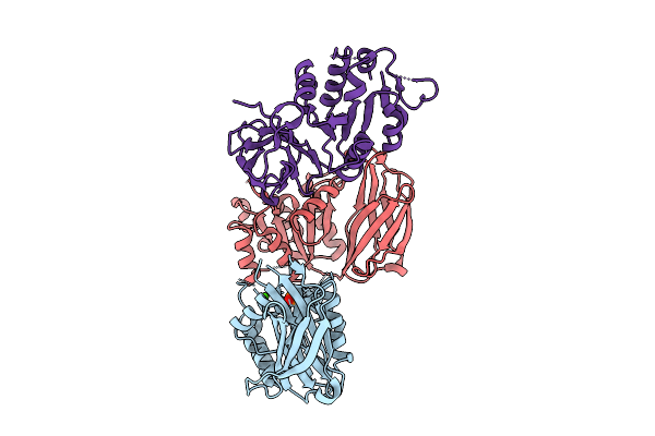
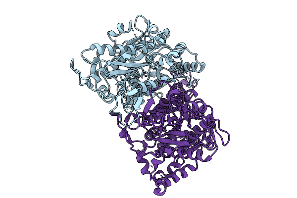
Structure Of The Sweet Receptor Bound To Sucralose In The Loose State, Extracellular Domain
Organism: Homo sapiens, Mus musculus
Method: ELECTRON MICROSCOPY Release Date: 2025-11-19 Classification: SIGNALING PROTEIN |
 |
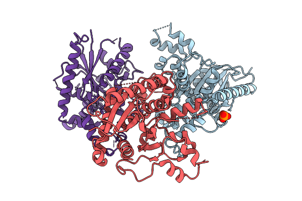
Open State Without Nuqm And Without Flavoprotein (Classification State 4) Of Pichia Pastoris Mitochondrial Complex I In Cmsp26 Nanodiscs
Organism: Komagataella pastoris
Method: ELECTRON MICROSCOPY Release Date: 2025-10-15 Classification: MEMBRANE PROTEIN Ligands: SF4, PLC, K, 3PE, CDL, NDP, ZN, EHZ |
 |
Open State Without Nuqm And With Flavoprotein (Classification State 3) Of Pichia Pastoris Mitochondrial Complex I In Cmsp26 Nanodiscs
Organism: Komagataella pastoris
Method: ELECTRON MICROSCOPY Release Date: 2025-10-15 Classification: MEMBRANE PROTEIN Ligands: SF4, PLC, FES, FMN, K, 3PE, CDL, NDP, ZN, EHZ |
 |
Closed State With Nuqm And Without Flavoprotein (Classification State 2) Of Pichia Pastoris Mitochondrial Complex I In Cmsp26 Nanodiscs
Organism: Komagataella pastoris
Method: ELECTRON MICROSCOPY Release Date: 2025-10-15 Classification: MEMBRANE PROTEIN Ligands: PLC, SF4, FES, K, 3PE, CDL, NDP, ZN, EHZ |
 |
Closed State With Nuqm And With Flavoprotein (Classification State 1) Of Pichia Pastoris Mitochondrial Complex I In Cmsp26 Nanodiscs
Organism: Komagataella pastoris
Method: ELECTRON MICROSCOPY Release Date: 2025-10-15 Classification: MEMBRANE PROTEIN Ligands: PLC, SF4, FES, FMN, K, 3PE, CDL, NDP, ZN, EHZ |
 |
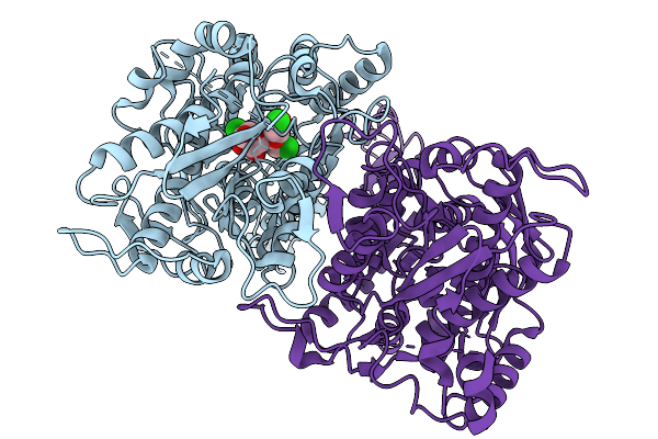
Crystal Structure Of Nir2 C-Terminal Domain In Complex With Phosphatidic Acid
Organism: Mus musculus
Method: X-RAY DIFFRACTION Release Date: 2025-10-15 Classification: LIPID BINDING PROTEIN Ligands: 44E, DW3, CA |
 |
Organism: Mus musculus
Method: X-RAY DIFFRACTION Release Date: 2025-10-15 Classification: LIPID BINDING PROTEIN Ligands: ZN |
 |
Organism: Homo sapiens
Method: X-RAY DIFFRACTION Release Date: 2025-10-15 Classification: LIPID BINDING PROTEIN Ligands: PO4 |
 |
Organism: Mus musculus
Method: X-RAY DIFFRACTION Release Date: 2025-10-15 Classification: LIPID BINDING PROTEIN Ligands: CA, PO4 |
 |
Organism: Mus musculus
Method: X-RAY DIFFRACTION Release Date: 2025-10-15 Classification: LIPID BINDING PROTEIN Ligands: PEG |
 |
Organism: Mus musculus
Method: SOLUTION NMR Release Date: 2025-09-03 Classification: CYTOSOLIC PROTEIN |
 |
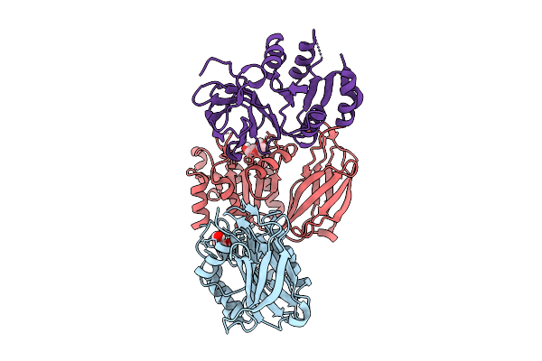
Organism: Homo sapiens, Mus musculus
Method: ELECTRON MICROSCOPY Release Date: 2025-09-03 Classification: SIGNALING PROTEIN |