
Search Count: 39
 |
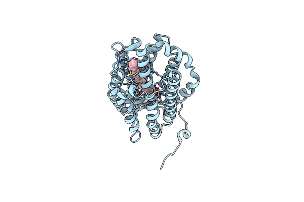
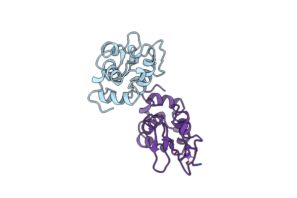
Crystal Structure Of Class Ie Ribonucleotide Reductase R2 Subunit Without Y150 Modification From Gardnerella Vaginalis
Organism: Gardnerella vaginalis atcc 14019
Method: X-RAY DIFFRACTION Resolution:1.90 Å Release Date: 2024-12-11 Classification: OXIDOREDUCTASE |
 |
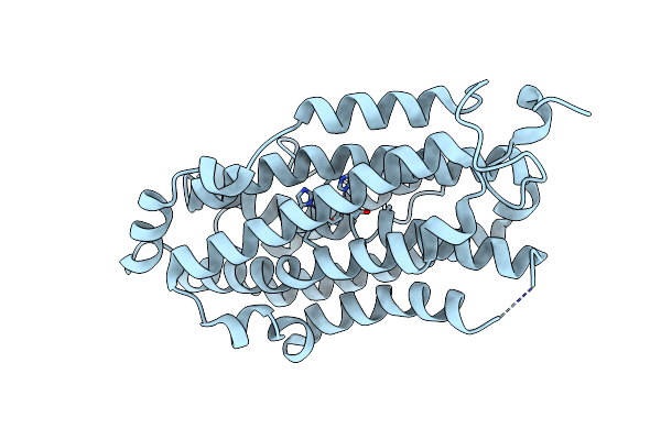
Crystal Structure Of Class Ie Ribonucleotide Reductase R2 Subunit With Post-Translational Modification Of Y150 Into A Dopa From Gardnerella Vaginalis
Organism: Gardnerella vaginalis atcc 14019
Method: X-RAY DIFFRACTION Release Date: 2024-12-11 Classification: OXIDOREDUCTASE |
 |
Ribonucleotide Reductase Class Ie R2 From Mesoplasma Florum, Catalytically Active Radical State Solved By Xfel
Organism: Mesoplasma florum l1
Method: X-RAY DIFFRACTION Release Date: 2023-11-01 Classification: OXIDOREDUCTASE |
 |
Ribonucleotide Reductase Class Ie R2 From Mesoplasma Florum, Radical-Lost Ground State
Organism: Mesoplasma florum l1
Method: X-RAY DIFFRACTION Release Date: 2023-11-01 Classification: OXIDOREDUCTASE Ligands: CA, GOL |
 |
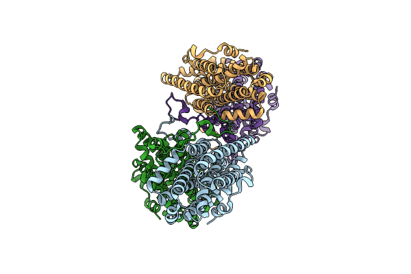
Xfel Structure Of Class Ib Ribonucleotide Reductase Dimanganese(Ii) Nrdf In Complex With Oxidized Nrdi From Bacillus Cereus
Organism: Bacillus cereus atcc 14579
Method: X-RAY DIFFRACTION Resolution:2.00 Å Release Date: 2022-09-21 Classification: OXIDOREDUCTASE Ligands: MN, UNX, FMN |
 |
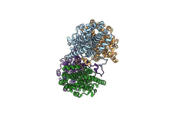
Xfel Structure Of Class Ib Ribonucleotide Reductase Dimanganese(Ii) Nrdf In Complex With Hydroquinone Nrdi From Bacillus Cereus
Organism: Bacillus cereus atcc 14579
Method: X-RAY DIFFRACTION Resolution:2.00 Å Release Date: 2022-09-21 Classification: OXIDOREDUCTASE Ligands: MN, FNR, UNX |
 |
Crystal Structure Of A Second Homolog Of R2-Like Ligand-Binding Oxidase In Sulfolobus Acidocaldarius (Sar2Loxii)
Organism: Sulfolobus acidocaldarius dsm 639
Method: X-RAY DIFFRACTION Resolution:2.26 Å Release Date: 2022-04-06 Classification: OXIDOREDUCTASE Ligands: MN3, FE |
 |
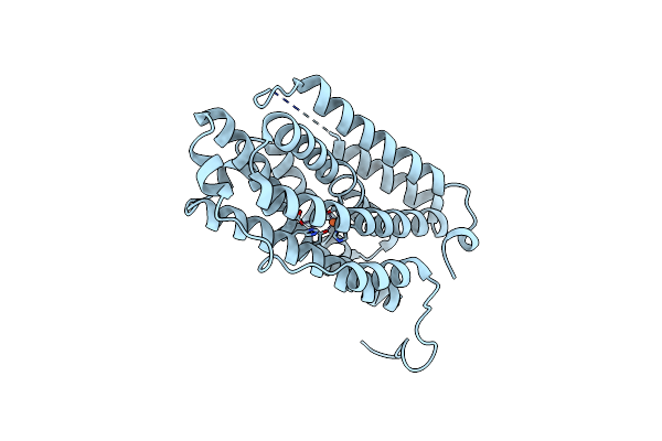
Crystal Structure Of R2-Like Ligand-Binding Oxidase From Saccharopolyspora Erythraea
Organism: Saccharopolyspora erythraea (strain atcc 11635 / dsm 40517 / jcm 4748 / nbrc 13426 / ncimb 8594 / nrrl 2338)
Method: X-RAY DIFFRACTION Resolution:1.38 Å Release Date: 2022-04-06 Classification: OXIDOREDUCTASE Ligands: MN3, FE, PLM, SO4 |
 |
Crystal Structure Of Ribonucleotide Reductase R2 Subunit Solved By Serial Synchrotron Crystallography
Organism: Saccharopolyspora erythraea (strain atcc 11635 / dsm 40517 / jcm 4748 / nbrc 13426 / ncimb 8594 / nrrl 2338)
Method: X-RAY DIFFRACTION Resolution:2.40 Å Release Date: 2020-10-07 Classification: OXIDOREDUCTASE Ligands: MN3, FE |
 |
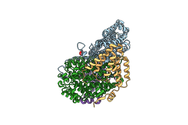
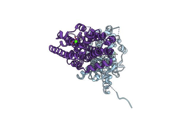
Xfel Structure Of The Soluble Methane Monooxygenase Hydroxylase And Regulatory Subunit Complex, From Methylosinus Trichosporium Ob3B, Diferric State
Organism: Methylosinus trichosporium ob3b
Method: X-RAY DIFFRACTION Resolution:1.95 Å Release Date: 2020-09-30 Classification: OXIDOREDUCTASE Ligands: GOL, FE |
 |
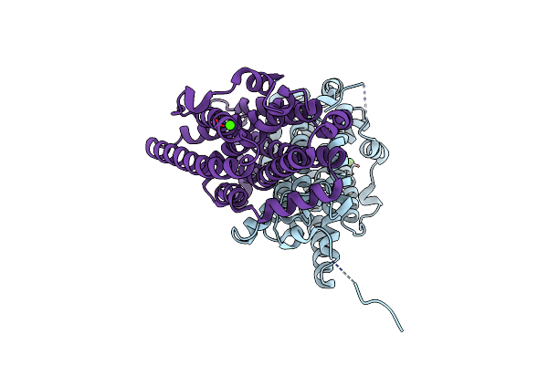
Xfel Structure Of The Soluble Methane Monooxygenase Hydroxylase And Regulatory Subunit Complex, From Methylosinus Trichosporium Ob3B, Diferrous State
Organism: Methylosinus trichosporium ob3b
Method: X-RAY DIFFRACTION Resolution:1.95 Å Release Date: 2020-09-30 Classification: OXIDOREDUCTASE Ligands: GOL, FE2 |
 |
Xfel Structure Of The Soluble Methane Monooxygenase Hydroxylase And Regulatory Subunit Complex, From Methylosinus Trichosporium Ob3B, Reoxidized Diferric State, 10S O2 Exposure.
Organism: Methylosinus trichosporium ob3b
Method: X-RAY DIFFRACTION Resolution:1.95 Å Release Date: 2020-09-30 Classification: OXIDOREDUCTASE Ligands: GOL, FE |
 |
Xfel Structure Of The Soluble Methane Monooxygenase Hydroxylase And Regulatory Subunit Complex, From Methylosinus Trichosporium Ob3B, T=0 Diferrous State Prior To Oxygen Activation
Organism: Methylosinus trichosporium ob3b
Method: X-RAY DIFFRACTION Resolution:2.00 Å Release Date: 2020-09-30 Classification: OXIDOREDUCTASE Ligands: GOL, FE2 |
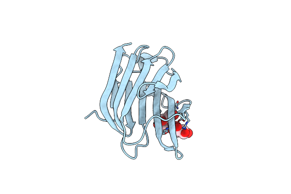 |
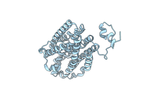
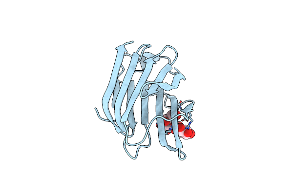
Structure Of Galectin-3C In Complex With Lactose Determined By Serial Crystallography Using A Silicon Nitride Membrane Support
Organism: Homo sapiens
Method: X-RAY DIFFRACTION Resolution:1.70 Å Release Date: 2020-07-29 Classification: SUGAR BINDING PROTEIN |
 |
Structure Of Galectin-3C In Complex With Lactose Determined By Serial Crystallography Using An Xtaltool Support
Organism: Homo sapiens
Method: X-RAY DIFFRACTION Resolution:1.70 Å Release Date: 2020-06-17 Classification: SUGAR BINDING PROTEIN Ligands: CL |
 |
Crystal Structure Of R2-Like Ligand-Binding Oxidase From Sulfolobus Acidocaldarius Solved By 3D Micro-Crystal Electron Diffraction
Organism: Sulfolobus acidocaldarius dsm 639
Method: ELECTRON CRYSTALLOGRAPHY Resolution:3.00 Å Release Date: 2019-04-24 Classification: OXIDOREDUCTASE Ligands: MN3, FE |
 |
Ribonucleotide Reductase Class Ie R2 From Mesoplasma Florum, Dopa-Active Form
Organism: Mesoplasma florum l1
Method: X-RAY DIFFRACTION Release Date: 2018-08-22 Classification: OXIDOREDUCTASE Ligands: CA |
 |
Organism: Mesoplasma florum (strain atcc 33453 / nbrc 100688 / nctc 11704 / l1)
Method: X-RAY DIFFRACTION Resolution:1.23 Å Release Date: 2018-08-22 Classification: OXIDOREDUCTASE Ligands: CA |
 |
A Rare Lysozyme Crystal Form Solved Using High-Redundancy 3D Electron Diffraction Data From Micron-Sized Needle Shaped Crystals
Organism: Gallus gallus
Method: ELECTRON CRYSTALLOGRAPHY Resolution:2.20 Å Release Date: 2018-03-28 Classification: HYDROLASE |
 |
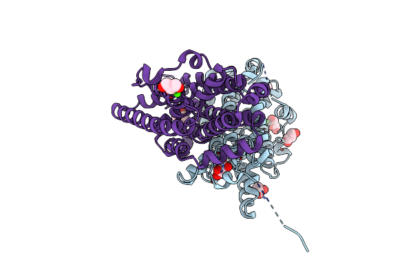
Structure Of Pas-Gaf Fragment Of Deinococcus Phytochrome By Serial Femtosecond Crystallography
Organism: Deinococcus radiodurans (strain atcc 13939 / dsm 20539 / jcm 16871 / lmg 4051 / nbrc 15346 / ncimb 9279 / r1 / vkm b-1422)
Method: X-RAY DIFFRACTION Resolution:1.65 Å Release Date: 2017-02-22 Classification: TRANSFERASE Ligands: LBV, NI, CL, EDO |