
Search Count: 24
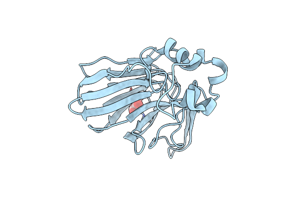 |
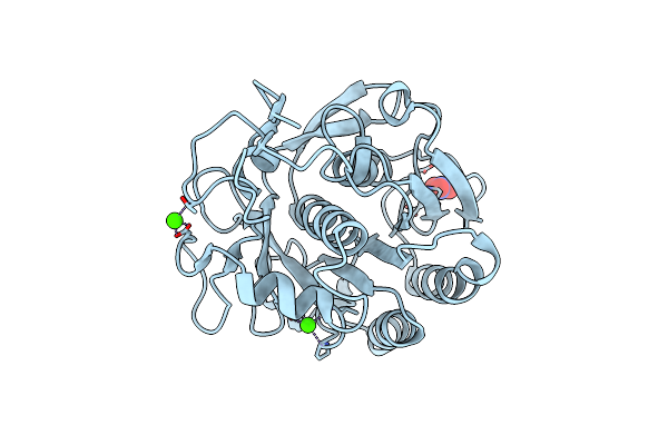
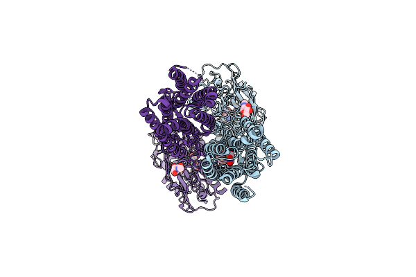
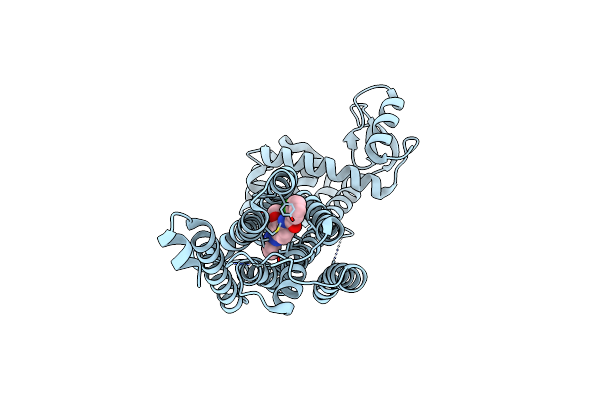
Serial Microseconds Crystallography At Id29 Using Sacla Extruder: Thaumatin
Organism: Thaumatococcus daniellii
Method: X-RAY DIFFRACTION Resolution:1.75 Å Release Date: 2025-01-22 Classification: PLANT PROTEIN Ligands: TLA |
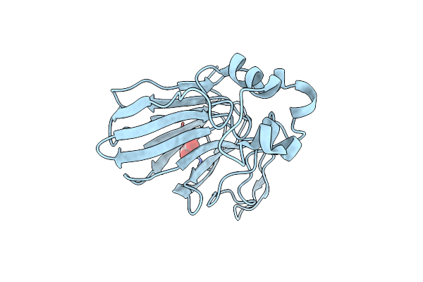 |
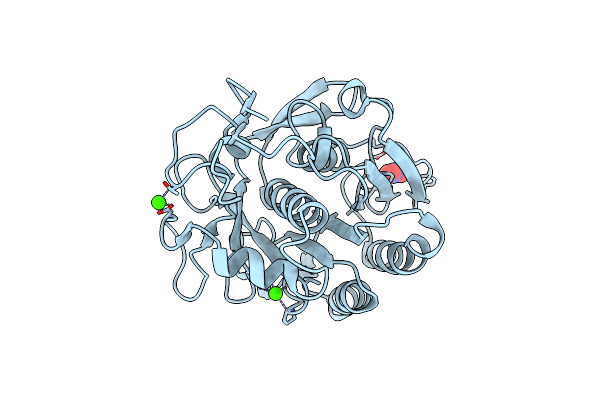
Organism: Thaumatococcus daniellii
Method: X-RAY DIFFRACTION Resolution:1.75 Å Release Date: 2025-01-22 Classification: PLANT PROTEIN Ligands: TLA |
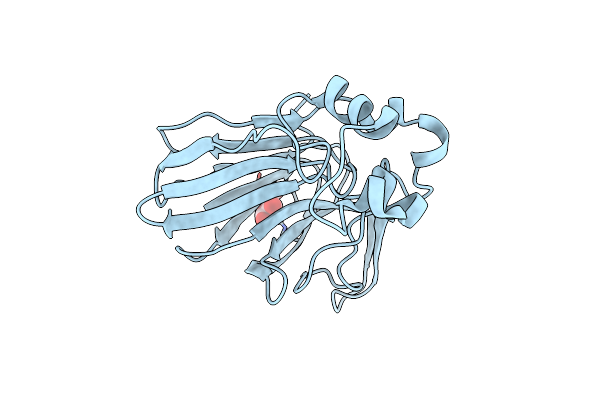 |
Organism: Thaumatococcus daniellii
Method: X-RAY DIFFRACTION Resolution:1.75 Å Release Date: 2025-01-22 Classification: PLANT PROTEIN Ligands: TLA |
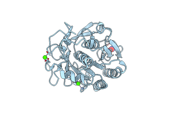 |
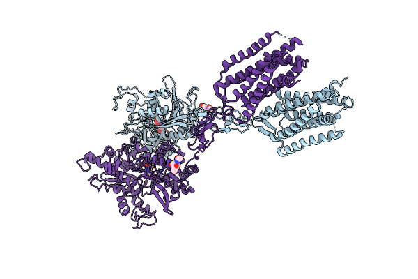
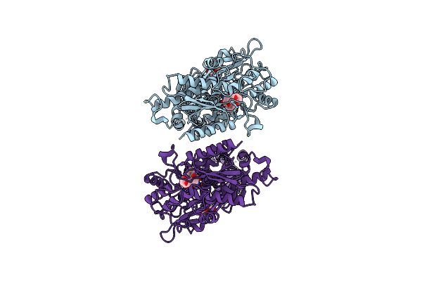
Serial Microseconds Crystallography At Id29 Using Fixed-Target (Small Foils): Proteinase K With 10 Um Spacing Between X-Ray Pulses
Organism: Parengyodontium album
Method: X-RAY DIFFRACTION Resolution:2.00 Å Release Date: 2025-01-22 Classification: HYDROLASE Ligands: CA, NO3 |
 |
Serial Microseconds Crystallography At Id29 Using Fixed-Target (Small Foils): Proteinase K With 20 Um Spacing Between X-Ray Pulses
Organism: Parengyodontium album
Method: X-RAY DIFFRACTION Resolution:2.00 Å Release Date: 2025-01-22 Classification: HYDROLASE Ligands: CA, NO3 |
 |
Serial Microseconds Crystallography At Id29 Using Fixed-Target (Large Foils): Proteinase K With 50 Um Spacing Between X-Ray Pulses
Organism: Parengyodontium album
Method: X-RAY DIFFRACTION Resolution:2.00 Å Release Date: 2025-01-22 Classification: HYDROLASE Ligands: CA, NO3 |
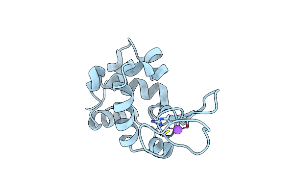 |
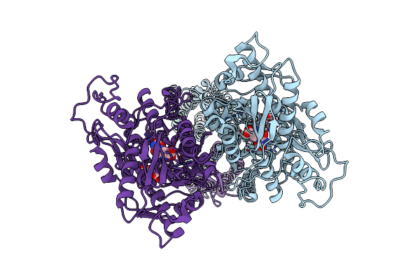
Serial Microseconds Crystallography At Id29 Using Fixed-Target (Si Chip): Lysozyme - Without Ligand Glcnac (Apo)
Organism: Gallus gallus
Method: X-RAY DIFFRACTION Resolution:2.05 Å Release Date: 2025-01-22 Classification: HYDROLASE |
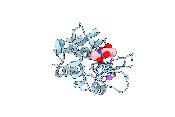 |
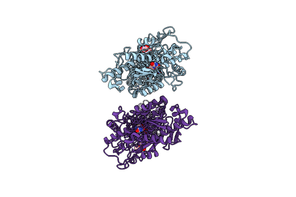
Serial Microseconds Crystallography At Id29 Using Fixed-Target (Si Chip): Lysozyme - With Ligand Glcnac
Organism: Gallus gallus
Method: X-RAY DIFFRACTION Resolution:2.05 Å Release Date: 2025-01-22 Classification: HYDROLASE |
 |
Serial Microseconds Crystallography At Id29 Using Fixed-Target (Small Foils): A2A Adenosine Receptor Co-Crystallised With Istradefylline
Organism: Homo sapiens
Method: X-RAY DIFFRACTION Resolution:2.50 Å Release Date: 2025-01-22 Classification: SIGNALING PROTEIN |
 |
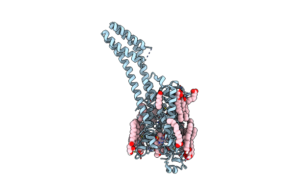
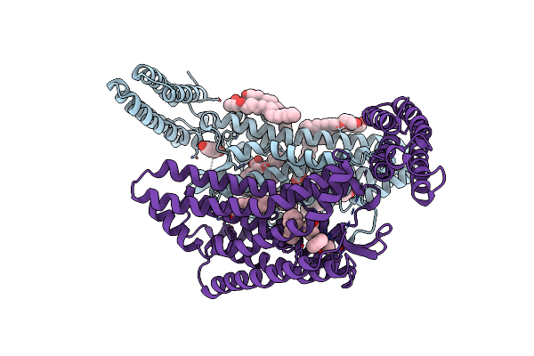
Human Fl Metabotropic Glutamate Receptor 5, Mglu5-5M With Quisqualate And Vu0424465
Organism: Homo sapiens
Method: ELECTRON MICROSCOPY Resolution:3.10 Å Release Date: 2024-11-06 Classification: MEMBRANE PROTEIN Ligands: NAG, QUS, XQT |
 |
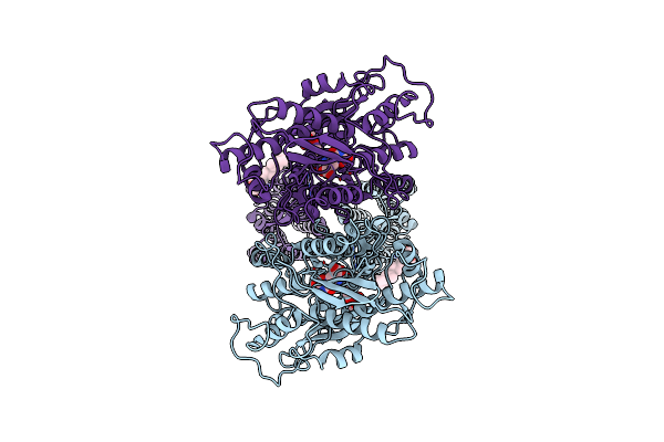
Human Fl Metabotropic Glutamate Receptor 5, Mglu5-5M With Quisqualate And Vu0424465, Conformer 1
Organism: Homo sapiens
Method: ELECTRON MICROSCOPY Resolution:3.20 Å Release Date: 2024-11-06 Classification: MEMBRANE PROTEIN Ligands: NAG, QUS, XQT |
 |
Human Fl Metabotropic Glutamate Receptor 5, Mglu5-5M With Quisqualate And Vu0424465, Conformer 2
Organism: Homo sapiens
Method: ELECTRON MICROSCOPY Resolution:3.50 Å Release Date: 2024-11-06 Classification: MEMBRANE PROTEIN Ligands: NAG, QUS, XQT |
 |
Human Fl Metabotropic Glutamate Receptor 5, Mglu5-5M With Agonist And Pam, W785A Mutant
Organism: Homo sapiens
Method: ELECTRON MICROSCOPY Resolution:3.40 Å Release Date: 2024-11-06 Classification: MEMBRANE PROTEIN Ligands: NAG, QUS |
 |
Human Fl Metabotropic Glutamate Receptor 5, Mglu5-5M With Quisqualate And Pam Vu29
Organism: Homo sapiens
Method: ELECTRON MICROSCOPY Release Date: 2024-11-06 Classification: MEMBRANE PROTEIN Ligands: NAG, QUS, XRQ |
 |
Human Fl Metabotropic Glutamate Receptor 5, Mglu5-5M With Quisqualate, Acc Conformation (Purified With Pam Vu0409551 But Not Modelled)
Organism: Homo sapiens
Method: ELECTRON MICROSCOPY Resolution:3.00 Å Release Date: 2024-11-06 Classification: MEMBRANE PROTEIN Ligands: NAG, QUS |
 |
Human Fl Metabotropic Glutamate Receptor 5, Mglu5-5M With Quisqualate, Rcc Conformation
Organism: Homo sapiens
Method: ELECTRON MICROSCOPY Release Date: 2024-11-06 Classification: MEMBRANE PROTEIN Ligands: NAG, QUS |
 |
Crystal Structure Of The Angiotensin Ii Type 2 Receptoror (At2R) In Complex With Ema401
Organism: Escherichia coli, Homo sapiens
Method: X-RAY DIFFRACTION Resolution:3.00 Å Release Date: 2022-02-09 Classification: MEMBRANE PROTEIN Ligands: VFD, OLC, OLA, FMT, HEZ |
 |
Organism: Homo sapiens
Method: ELECTRON MICROSCOPY Release Date: 2021-09-08 Classification: MEMBRANE PROTEIN Ligands: NAG, QUS, Y01 |
 |
Thermostabilised Full Length Human Mglur5-5M With Orthosteric Antagonist, Ly341495
Organism: Homo sapiens
Method: ELECTRON MICROSCOPY Release Date: 2021-09-08 Classification: MEMBRANE PROTEIN Ligands: NAG, Z99 |
 |
Thermostabilised 7Tm Domain Of Human Mglu5 Receptor Bound To Photoswitchable Ligand Alloswitch-1
Organism: Homo sapiens, Enterobacteria phage t4
Method: X-RAY DIFFRACTION Resolution:2.54 Å Release Date: 2021-09-08 Classification: SIGNALING PROTEIN Ligands: 4YI |