
Search Count: 29
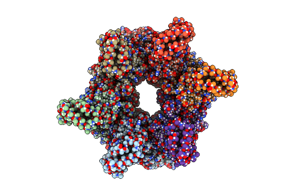 |
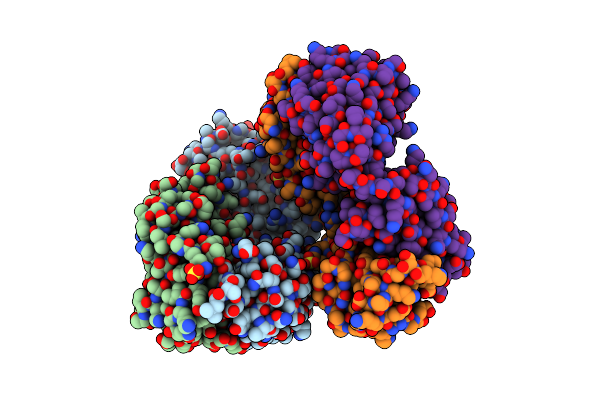
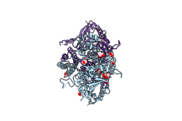
Organism: Caulobacter vibrioides na1000
Method: ELECTRON MICROSCOPY Release Date: 2024-08-14 Classification: MOTOR PROTEIN Ligands: ADP, MG, EPE |
 |
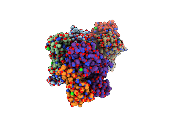
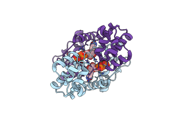
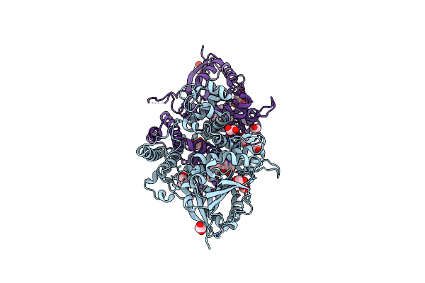
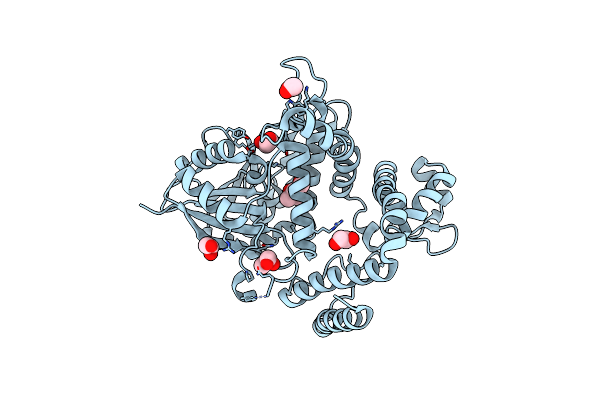
Organism: Caulobacter vibrioides na1000
Method: ELECTRON MICROSCOPY Release Date: 2024-08-14 Classification: MOTOR PROTEIN Ligands: ADP, EPE, MG, ANP |
 |
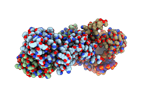
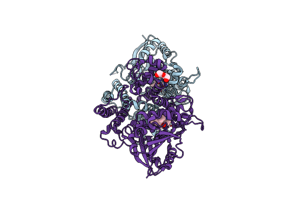
Organism: Caulobacter vibrioides na1000
Method: ELECTRON MICROSCOPY Release Date: 2024-08-14 Classification: MOTOR PROTEIN Ligands: EPE |
 |
Crystal Structure Of The C-Terminally Truncated Transcriptional Repressor Protein Korb From The Rk2 Plasmid Complexed With Ctp-Gamma-S
Organism: Escherichia coli
Method: X-RAY DIFFRACTION Resolution:2.30 Å Release Date: 2024-02-21 Classification: DNA BINDING PROTEIN Ligands: CTP, CL |
 |
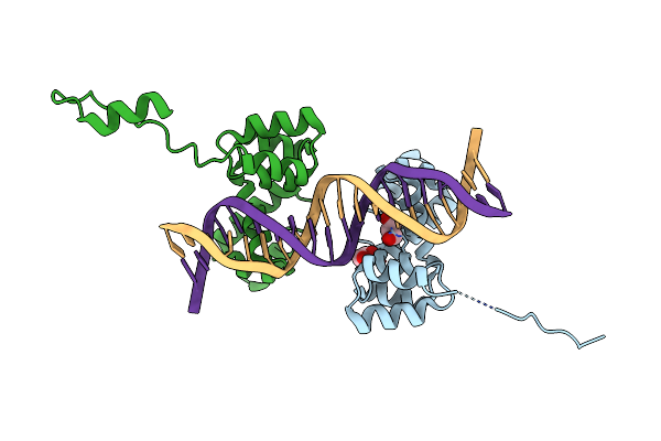
Crystal Structure Of The Rk2 Plasmid Encoded Co-Complex Of The C-Terminally Truncated Transcriptional Repressor Protein Korb Complexed With The Partner Repressor Protein Kora Bound To Oa-Dna
Organism: Escherichia coli
Method: X-RAY DIFFRACTION Resolution:2.70 Å Release Date: 2024-02-21 Classification: DNA BINDING PROTEIN Ligands: SO4 |
 |
Crystal Structure Of C-Terminally Truncated Bacillus Subtilis Nucleoid Occlusion Protein (Noc) Complexed To The Noc-Binding Site (Nbs)
Organism: Bacillus subtilis (strain 168), Synthetic construct
Method: X-RAY DIFFRACTION Resolution:2.90 Å Release Date: 2022-03-09 Classification: DNA BINDING PROTEIN |
 |
Organism: Oryctolagus cuniculus, Homo sapiens
Method: X-RAY DIFFRACTION Resolution:2.85 Å Release Date: 2021-06-16 Classification: IMMUNE SYSTEM Ligands: SO4 |
 |
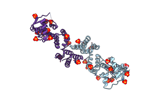
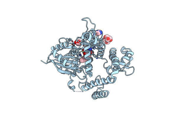
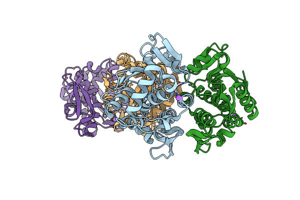
Crystal Structure Of The C-Terminally Truncated Chromosome-Partitioning Protein Parb From Caulobacter Crescentus Complexed With Ctp-Gamma-S
Organism: Caulobacter vibrioides (strain na1000 / cb15n)
Method: X-RAY DIFFRACTION Resolution:2.73 Å Release Date: 2021-04-28 Classification: DNA BINDING PROTEIN Ligands: CTP, MG |
 |
Crystal Structure Of C-Terminally Truncated Geobacillus Thermoleovorans Nucleoid Occlusion Protein Noc
Organism: Geobacillus thermoleovorans ccb_us3_uf5
Method: X-RAY DIFFRACTION Resolution:2.50 Å Release Date: 2021-02-17 Classification: DNA BINDING PROTEIN Ligands: SO4, GOL |
 |
Crystal Structure Of N- And C-Terminally Truncated Geobacillus Thermoleovorans Nucleoid Occlusion Protein Noc
Organism: Geobacillus thermoleovorans ccb_us3_uf5
Method: X-RAY DIFFRACTION Resolution:2.95 Å Release Date: 2021-02-17 Classification: DNA BINDING PROTEIN Ligands: SO4 |
 |
Crystal Structure Of The C-Terminally Truncated Chromosome-Partitioning Protein Parb From Caulobacter Crescentus Complexed To The Centromeric Pars Site
Organism: Caulobacter vibrioides (strain na1000 / cb15n), Caulobacter vibrioides
Method: X-RAY DIFFRACTION Resolution:2.90 Å Release Date: 2020-10-14 Classification: DNA BINDING PROTEIN |
 |
Organism: Aspergillus bombycis
Method: X-RAY DIFFRACTION Resolution:1.98 Å Release Date: 2020-10-14 Classification: BIOSYNTHETIC PROTEIN Ligands: EDO, GOL, NA |
 |
Organism: Aspergillus bombycis
Method: X-RAY DIFFRACTION Resolution:1.99 Å Release Date: 2020-10-14 Classification: BIOSYNTHETIC PROTEIN Ligands: F56, EDO, GOL |
 |
Organism: Aspergillus bombycis
Method: X-RAY DIFFRACTION Resolution:2.40 Å Release Date: 2020-10-14 Classification: BIOSYNTHETIC PROTEIN Ligands: F5F, PEG |
 |
Organism: Hymenoscyphus scutula
Method: X-RAY DIFFRACTION Resolution:1.53 Å Release Date: 2020-10-14 Classification: BIOSYNTHETIC PROTEIN Ligands: F56, IMD, GOL, B3P |
 |
Organism: Hymenoscyphus scutula
Method: X-RAY DIFFRACTION Resolution:1.33 Å Release Date: 2020-10-14 Classification: BIOSYNTHETIC PROTEIN Ligands: MPD, EDO, GOL |
 |
Crystal Structure Of The Dna-Binding Domain Of The Nucleoid Occlusion Factor (Noc) Complexed To The Noc-Binding Site (Nbs)
Organism: Bacillus subtilis (strain 168), Synthetic construct
Method: X-RAY DIFFRACTION Resolution:2.23 Å Release Date: 2020-08-05 Classification: DNA BINDING PROTEIN |
 |
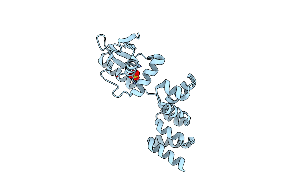
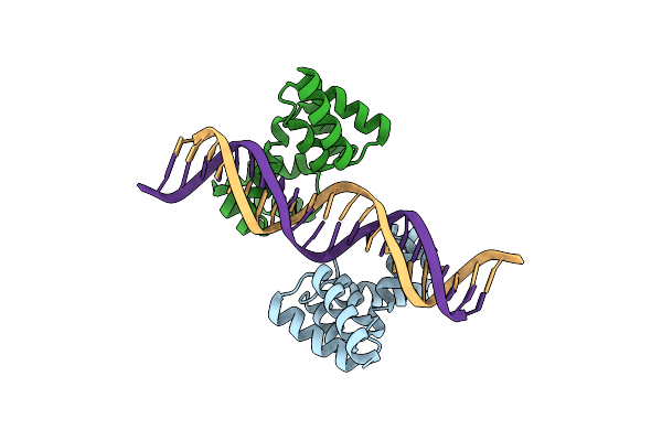
Crystal Structure Of The Dna Binding Domain Of The Chromosome-Partitioning Protein Parb Complexed To The Centromeric Pars Site
Organism: Caulobacter vibrioides na1000, Caulobacter vibrioides
Method: X-RAY DIFFRACTION Resolution:2.40 Å Release Date: 2020-07-15 Classification: DNA BINDING PROTEIN Ligands: GOL |
 |
Crystal Structure Of Type-I Ribosome-Inactivating Protein Trichobakin (Tbk)
Organism: Trichosanthes sp. bac kan 8-98
Method: X-RAY DIFFRACTION Resolution:2.00 Å Release Date: 2020-05-27 Classification: HYDROLASE |
 |
Solution Nmr Structure Of Type-I Ribosome-Inactivating Protein Trichobakin (Tbk)
Organism: Trichosanthes sp. bac kan 8-98
Method: SOLUTION NMR Release Date: 2020-05-06 Classification: HYDROLASE |