
Search Count: 35
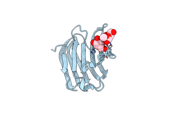 |
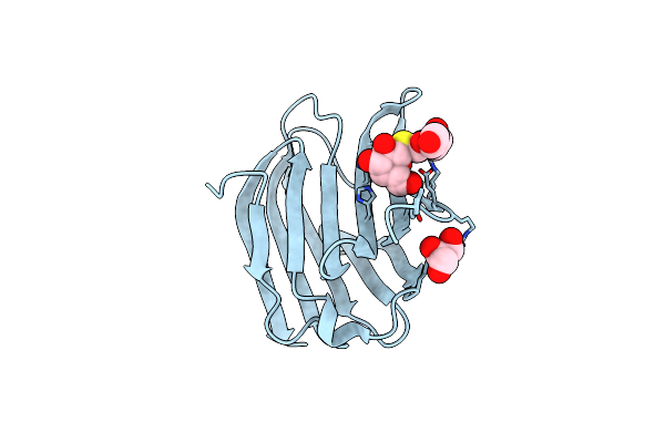
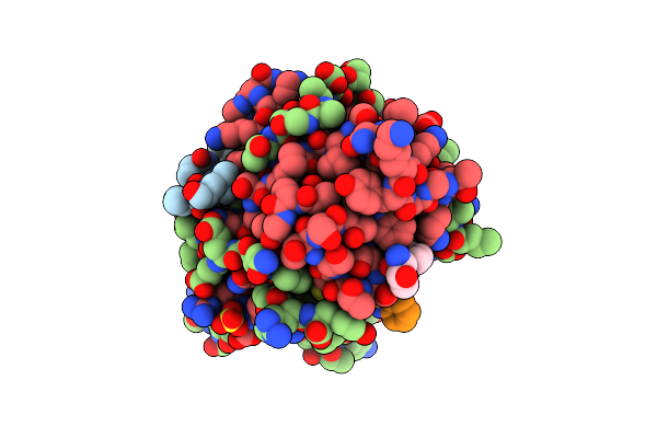
Galectin-3 In Complex With Methyl 2,6-Anhydro-3-Deoxy-3-S-(B-D-Galactopyranosyl)-3-Thio-D-Glycero-L-Altro-Heptonate
Organism: Homo sapiens
Method: X-RAY DIFFRACTION Resolution:1.75 Å Release Date: 2023-09-13 Classification: SUGAR BINDING PROTEIN Ligands: CL, YIO, VPH |
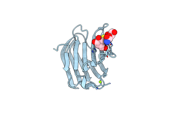 |
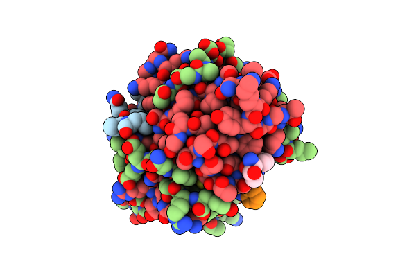
Galectin-3 In Complex With 2,6-Anhydro-3-Deoxy-3-S-(Beta-D-Galactopyranosyl)-3-Thio-D-Glycero-L-Altro-Heptonamide
Organism: Homo sapiens
Method: X-RAY DIFFRACTION Resolution:1.80 Å Release Date: 2023-09-13 Classification: SUGAR BINDING PROTEIN Ligands: YIO, VPL, MG |
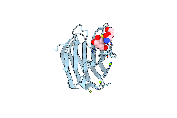 |
Galectin-3 In Complex With 2,6-Anhydro-3-Deoxy-3-S-(Beta-D-Galactopyranosyl)-3-Thio-D-Glycero-D-Galacto-Heptonamide
Organism: Homo sapiens
Method: X-RAY DIFFRACTION Resolution:1.80 Å Release Date: 2023-09-13 Classification: SUGAR BINDING PROTEIN Ligands: VPQ, YIO, MG |
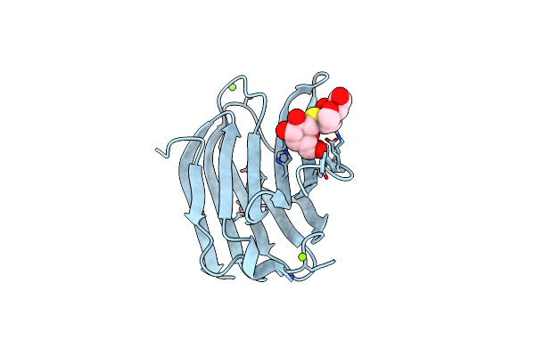 |
Galectin-3 In Complex With 2,6-Anhydro-5-S-(Beta-D-Galactopyranosyl)-5-Thio-D-Altritol
Organism: Homo sapiens
Method: X-RAY DIFFRACTION Resolution:1.80 Å Release Date: 2023-09-13 Classification: SUGAR BINDING PROTEIN Ligands: MG, AH2, YIO |
 |
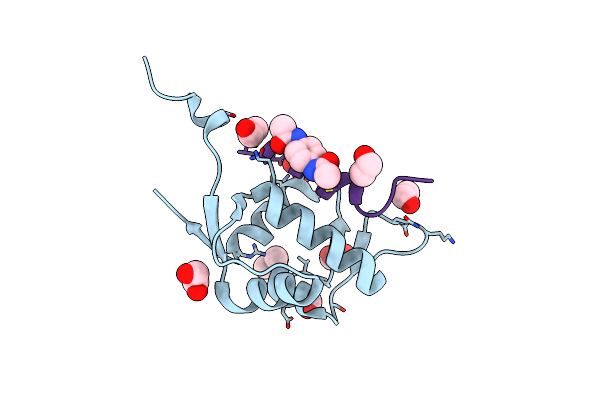
Galectin-3 Carbohydrate Recognition Domain In Complex With Thiodigalactoside At 1.8 Resolution
Organism: Homo sapiens
Method: X-RAY DIFFRACTION Resolution:1.80 Å Release Date: 2023-09-13 Classification: SUGAR BINDING PROTEIN Ligands: CL, GOL |
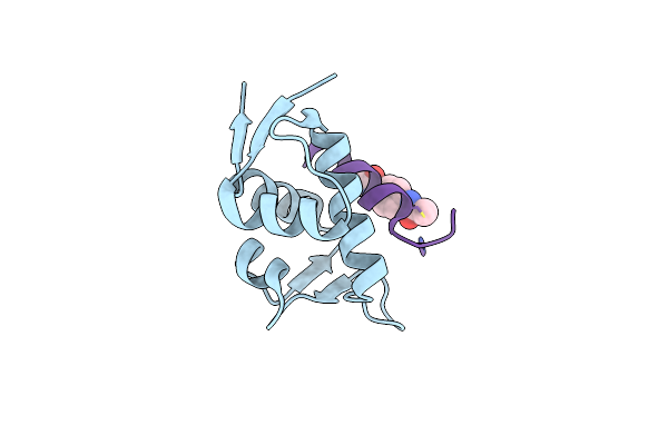 |
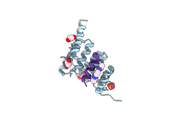
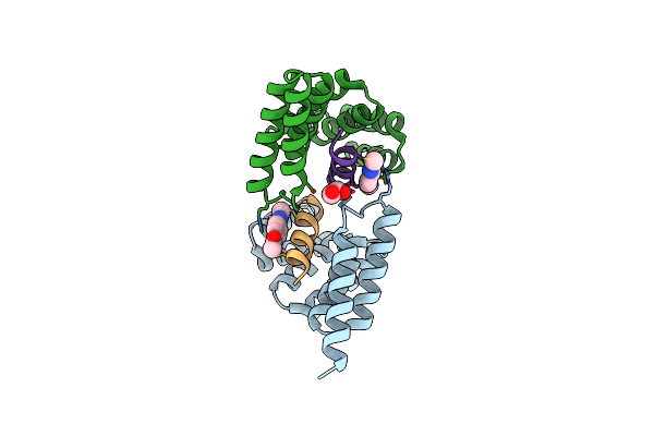
Structure Of The Mdm2 P53 Binding Domain In Complex With H101, An All-D Helicon Polypeptide
Organism: Homo sapiens, Synthetic construct
Method: X-RAY DIFFRACTION Resolution:1.61 Å Release Date: 2023-02-15 Classification: LIGASE Ligands: CL, WHL |
 |
Structure Of The Mdm2 P53 Binding Domain In Complex With H102, An All-D Helicon Polypeptide
Organism: Homo sapiens, Synthetic construct
Method: X-RAY DIFFRACTION Resolution:1.28 Å Release Date: 2023-02-15 Classification: LIGASE Ligands: EDO, CL, GOL, WHL, IMD |
 |
Structure Of The Mdm2 P53 Binding Domain In Complex With H103, An All-D Helicon Polypeptide
Organism: Homo sapiens, Synthetic construct
Method: X-RAY DIFFRACTION Resolution:1.86 Å Release Date: 2023-02-15 Classification: LIGASE Ligands: SO4, CL, WHL |
 |
Structure Of The Mdm2 P53 Binding Domain In Complex With H103, An All-D Helicon Polypeptide, Alternative C-Terminus
Organism: Homo sapiens, Synthetic construct
Method: X-RAY DIFFRACTION Resolution:1.40 Å Release Date: 2023-02-15 Classification: LIGASE Ligands: EDO, SO4, DMS, WHL |
 |
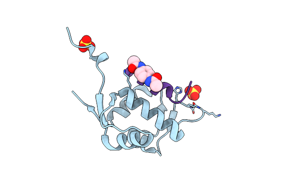
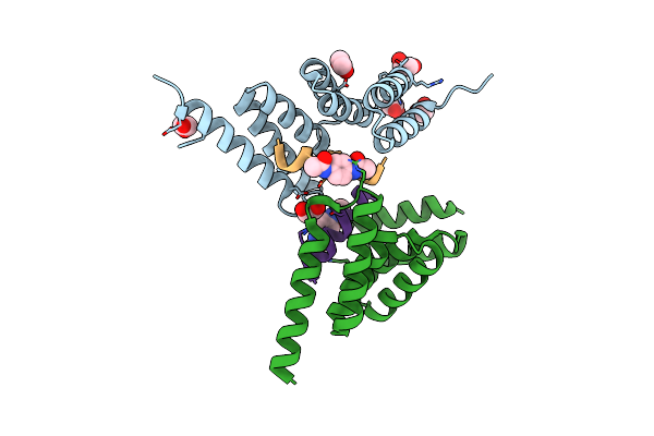
Structure Of The Stub1 Tpr Domain In Complex With H201, An All-D Helicon Polypeptide
Organism: Homo sapiens, Synthetic construct
Method: X-RAY DIFFRACTION Resolution:1.69 Å Release Date: 2023-02-15 Classification: LIGASE Ligands: SO4, EDO, WHL |
 |
Structure Of The Stub1 Tpr Domain In Complex With H202, An All-D Helicon Polypeptide
Organism: Homo sapiens, Synthetic construct
Method: X-RAY DIFFRACTION Resolution:1.73 Å Release Date: 2023-02-15 Classification: LIGASE Ligands: EDO, WHL |
 |
Structure Of The Stub1 Tpr Domain In Complex With H203, An All-D Helicon Polypeptide
Organism: Homo sapiens, Synthetic construct
Method: X-RAY DIFFRACTION Resolution:1.56 Å Release Date: 2023-02-15 Classification: LIGASE Ligands: EDO, WHL |
 |
Structure Of The Stub1 Tpr Domain In Complex With H204, An All-D Helicon Polypeptide
Organism: Homo sapiens, Synthetic construct
Method: X-RAY DIFFRACTION Resolution:2.21 Å Release Date: 2023-02-15 Classification: LIGASE Ligands: WHL, EDO |
 |
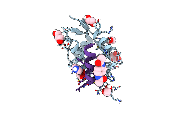
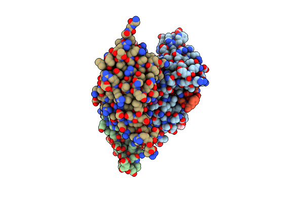
Crystal Structure Of Deuterated Gamma-Chymotrypsin At Ph 7.5, Room Temperature
Organism: Bos taurus
Method: X-RAY DIFFRACTION Resolution:1.05 Å Release Date: 2021-09-01 Classification: HYDROLASE Ligands: IOD |
 |
Crystal Structure Of Deuterated Gamma-Chymotrypsin At Ph 7.5, Cryo Temperature
Organism: Bos taurus
Method: X-RAY DIFFRACTION Resolution:1.00 Å Release Date: 2021-09-01 Classification: HYDROLASE Ligands: MLA, IOD |
 |
Organism: Bos taurus
Method: X-RAY DIFFRACTION Resolution:1.05 Å Release Date: 2021-09-01 Classification: HYDROLASE Ligands: IOD |
 |
Organism: Bos taurus
Method: X-RAY DIFFRACTION Resolution:1.05 Å Release Date: 2021-09-01 Classification: HYDROLASE Ligands: MLI, IOD |
 |
Crystal Structure Of Deuterated Gamma-Chymotrypsin At Ph 5.6, Room Temperature
Organism: Bos taurus
Method: X-RAY DIFFRACTION Resolution:1.05 Å Release Date: 2021-09-01 Classification: HYDROLASE Ligands: IOD, SO4 |
 |
Crystal Structure Of Deuterated Gamma-Chymotrypsin At Ph 5.6, Cryo Temperature
Organism: Bos taurus
Method: X-RAY DIFFRACTION Resolution:1.10 Å Release Date: 2021-09-01 Classification: HYDROLASE Ligands: MLA, IOD |
 |
Organism: Bos taurus
Method: X-RAY DIFFRACTION Resolution:1.05 Å Release Date: 2021-09-01 Classification: HYDROLASE Ligands: IOD, SO4 |