
Search Count: 30
 |
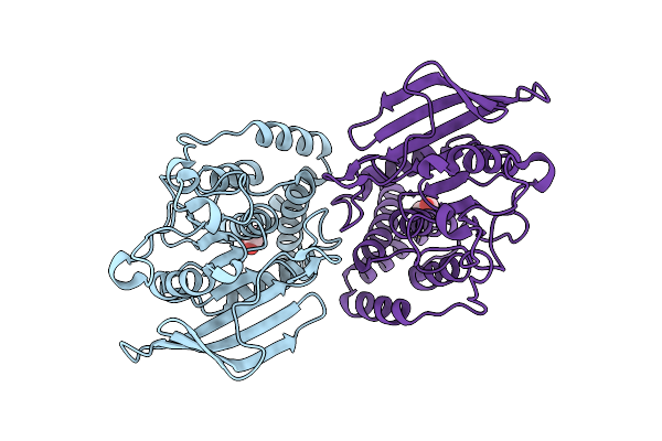
Organism: Homo sapiens
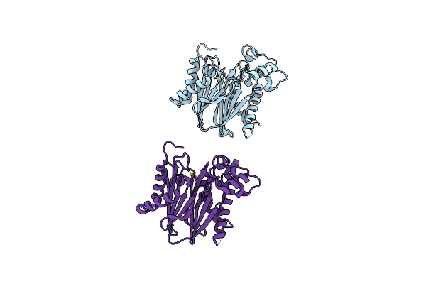
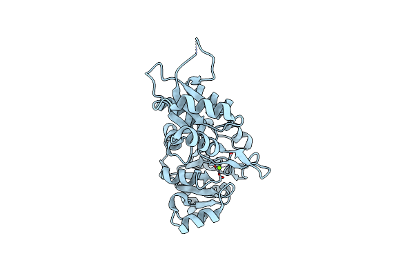
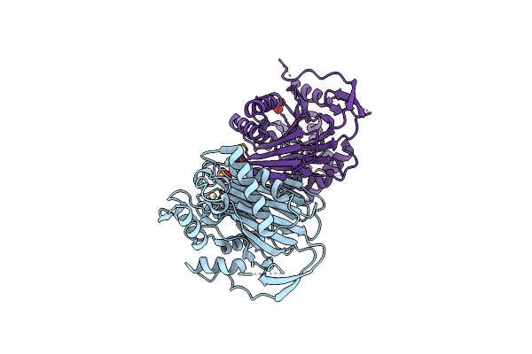
Method: ELECTRON MICROSCOPY Resolution:3.90 Å Release Date: 2025-04-16 Classification: CELL CYCLE Ligands: ZN |
 |
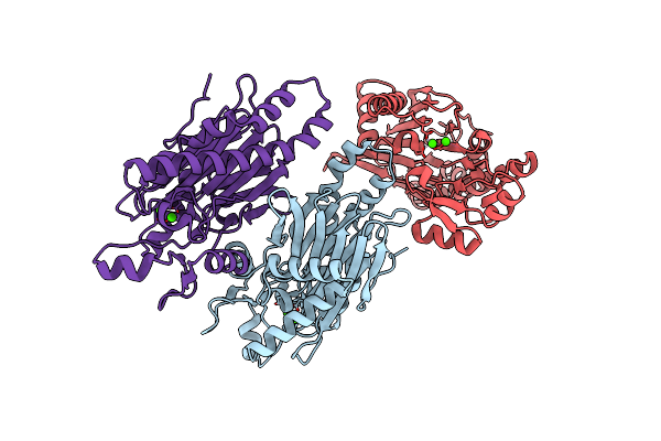
Organism: Homo sapiens
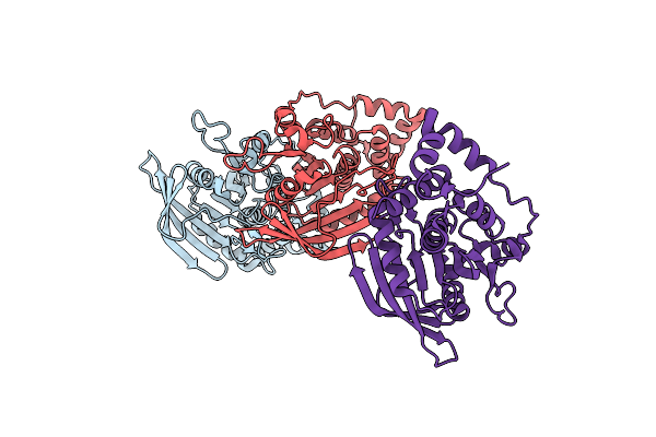
Method: ELECTRON MICROSCOPY Resolution:3.90 Å Release Date: 2025-04-16 Classification: CELL CYCLE Ligands: ZN |
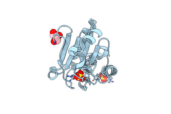 |
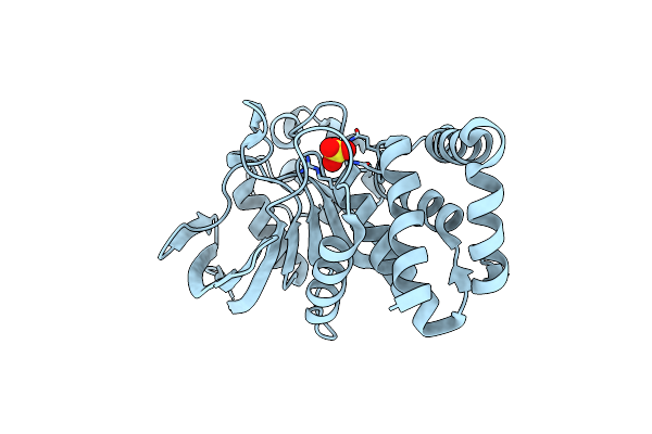
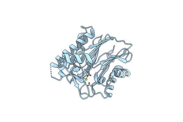
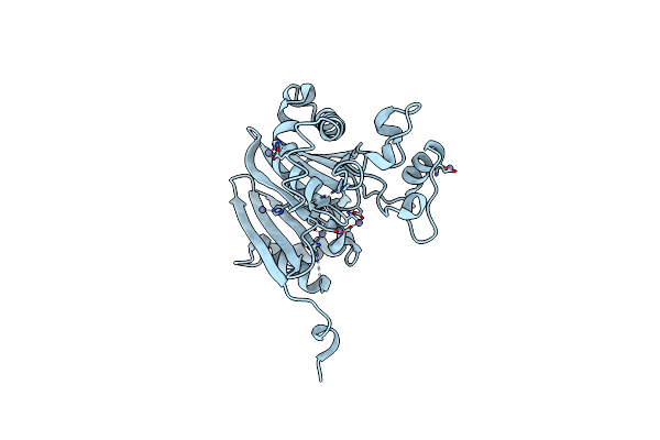
Crystal Structure Of Human Tyrosine Phosphatase-Like Serine/Threonine/Tyrosine-Interacting Protein
Organism: Homo sapiens
Method: X-RAY DIFFRACTION Resolution:1.60 Å Release Date: 2007-08-28 Classification: HYDROLASE Ligands: SO4, GOL |
 |
Organism: Trypanosoma brucei
Method: X-RAY DIFFRACTION Resolution:2.05 Å Release Date: 2007-07-24 Classification: HYDROLASE Ligands: MN, PO4 |
 |
Crystal Structure Of Human Carboxy-Terminal Domain Rna Polymerase Ii Polypeptide A Small Phosphatase 2
Organism: Homo sapiens
Method: X-RAY DIFFRACTION Resolution:2.51 Å Release Date: 2007-06-19 Classification: HYDROLASE Ligands: MG |
 |
Organism: Homo sapiens
Method: X-RAY DIFFRACTION Resolution:2.25 Å Release Date: 2007-04-03 Classification: HYDROLASE Ligands: CA, PLP |
 |
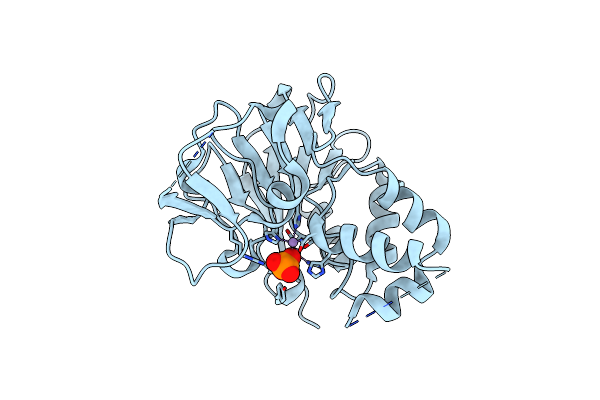
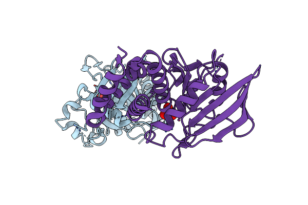
Crystal Structure Of The Serine/Threonine Phosphatase Domain Of Human Ppm1B
Organism: Homo sapiens
Method: X-RAY DIFFRACTION Resolution:1.82 Å Release Date: 2007-04-03 Classification: HYDROLASE Ligands: MG |
 |
Organism: Homo sapiens
Method: X-RAY DIFFRACTION Resolution:1.70 Å Release Date: 2007-04-03 Classification: HYDROLASE Ligands: SO4 |
 |
Organism: Mus musculus
Method: X-RAY DIFFRACTION Resolution:1.90 Å Release Date: 2007-03-20 Classification: HYDROLASE Ligands: PO4 |
 |
Organism: Homo sapiens
Method: X-RAY DIFFRACTION Resolution:1.72 Å Release Date: 2007-03-13 Classification: HYDROLASE |
 |
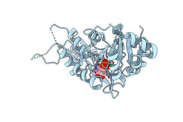
Crystal Structure Of Human Pyridoxal Phosphate Phosphatase With Mg2+ At 1.9 A Resolution
Organism: Homo sapiens
Method: X-RAY DIFFRACTION Resolution:1.90 Å Release Date: 2007-03-13 Classification: HYDROLASE Ligands: MG |
 |
Crystal Structure Of A C-Terminal Phosphatase Domain Of Rattus Norvegicus Ortholog Of Human Protein Tyrosine Phosphatase, Receptor Type, D (Ptprd)
Organism: Rattus norvegicus
Method: X-RAY DIFFRACTION Resolution:2.00 Å Release Date: 2006-11-21 Classification: HYDROLASE |
 |
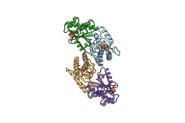
Crystal Structure Of Mitogen-Activated Protein Kinase Kinase Kinase 7 Interacting Protein 1 From Anopheles Gambiae
Organism: Anopheles gambiae
Method: X-RAY DIFFRACTION Resolution:3.00 Å Release Date: 2006-11-14 Classification: TRANSFERASE |
 |
Organism: Homo sapiens
Method: X-RAY DIFFRACTION Resolution:2.25 Å Release Date: 2006-11-07 Classification: HYDROLASE Ligands: MG |
 |
Crystal Structure Of A Phosphatase From A Pathogenic Strain Toxoplasma Gondii
Organism: Toxoplasma gondii
Method: X-RAY DIFFRACTION Resolution:1.90 Å Release Date: 2006-10-31 Classification: HYDROLASE Ligands: SO4, PR |
 |
Crystal Structure Of Anopheles Gambiae Ser/Thr Phosphatase Complexed With Zn2+
Organism: Anopheles gambiae
Method: X-RAY DIFFRACTION Resolution:1.70 Å Release Date: 2006-10-24 Classification: HYDROLASE Ligands: ZN |
 |
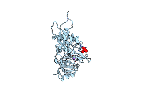
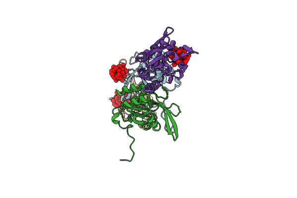
Crystal Structure Of The Human Tyrosine Receptor Phosphate Gamma In Complex With Vanadate
Organism: Homo sapiens
Method: X-RAY DIFFRACTION Resolution:2.60 Å Release Date: 2006-09-05 Classification: HYDROLASE Ligands: VO4 |
 |
Organism: Homo sapiens
Method: X-RAY DIFFRACTION Resolution:2.10 Å Release Date: 2006-08-29 Classification: HYDROLASE Ligands: KEG |
 |
Organism: Homo sapiens
Method: X-RAY DIFFRACTION Resolution:2.23 Å Release Date: 2006-08-29 Classification: HYDROLASE Ligands: GOL |
 |
Crystal Structure Of Serine-Threonine Phosphatase 2C From Toxoplasma Gondii
Organism: Toxoplasma gondii
Method: X-RAY DIFFRACTION Resolution:2.04 Å Release Date: 2006-08-29 Classification: HYDROLASE Ligands: CA |