
Search Count: 20
 |
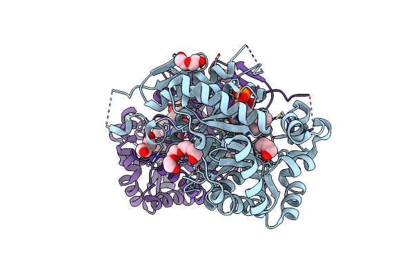
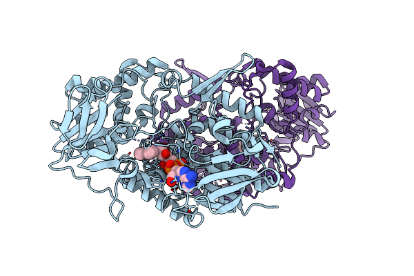
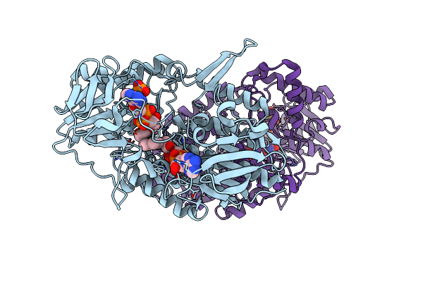
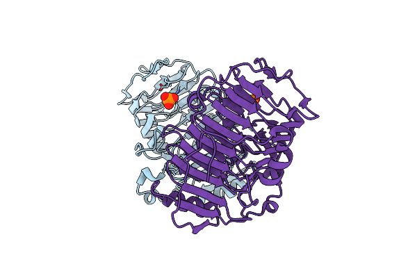
Type Ii Baeyer-Villiger Monooxygenase.The Oxygenating Constituent Of 3,6-Diketocamphane Monooxygenase From Cam Plasmid Of Pseudomonas Putida In Complex With Fmn.
Organism: Pseudomonas putida
Method: X-RAY DIFFRACTION Resolution:1.93 Å Release Date: 2015-09-09 Classification: OXIDOREDUCTASE Ligands: PIN, GOL, SO4, CL |
 |
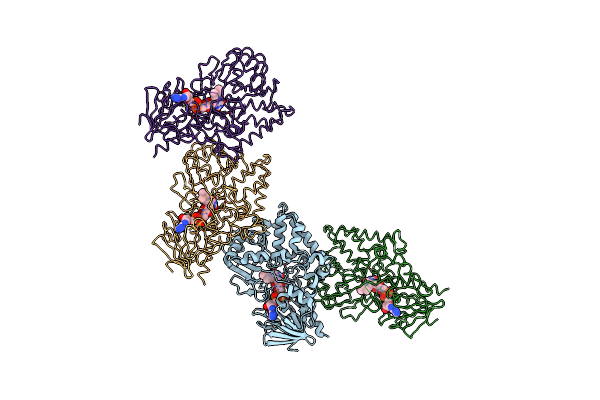
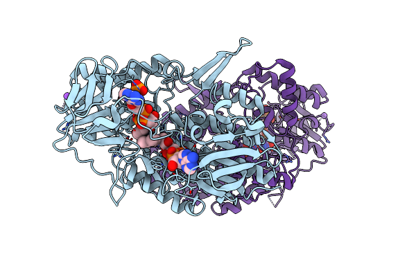
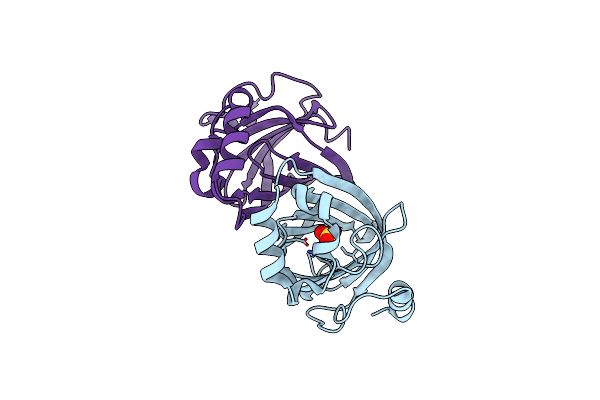
Type Ii Baeyer-Villiger Monooxygenase.The Oxygenating Constituent Of 3,6-Diketocamphane Monooxygenase From Cam Plasmid Of Pseudomonas Putida In Complex With Fmn.
Organism: Pseudomonas putida
Method: X-RAY DIFFRACTION Resolution:1.90 Å Release Date: 2015-08-26 Classification: OXIDOREDUCTASE Ligands: FMN, PG4, PEG, PGE |
 |
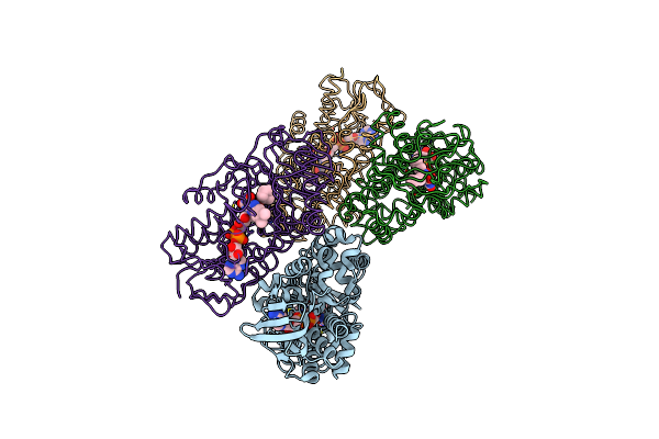
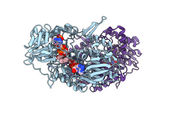
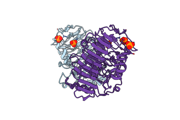
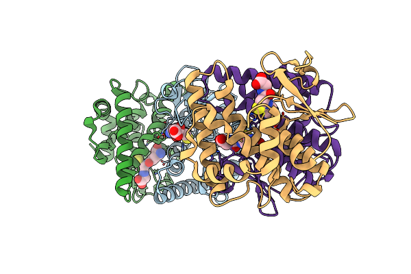
Epsilon-Caprolactone-Bound Crystal Structure Of Cyclohexanone Monooxygenase In The Tight Conformation
Organism: Rhodococcus sp. hi-31
Method: X-RAY DIFFRACTION Resolution:1.94 Å Release Date: 2014-10-15 Classification: OXIDOREDUCTASE Ligands: FAD, NAP, ECE, BCN |
 |
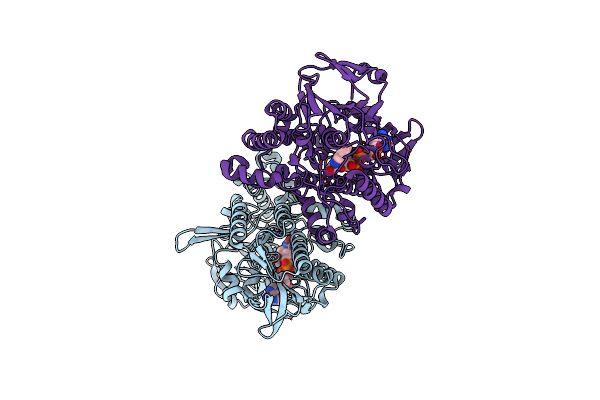
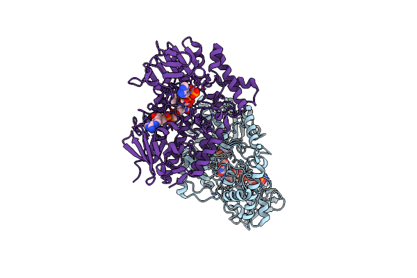
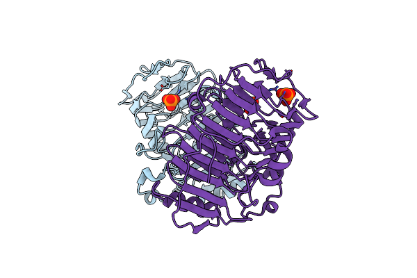
Epsilon-Caprolactone-Bound Crystal Structure Of Cyclohexanone Monooxygenase In The Loose Conformation
Organism: Rhodococcus sp. hi-31
Method: X-RAY DIFFRACTION Resolution:2.51 Å Release Date: 2014-10-15 Classification: OXIDOREDUCTASE Ligands: FAD, NAP, ECE, PTD |
 |
Organism: Brevibacterium oxydans
Method: X-RAY DIFFRACTION Resolution:3.00 Å Release Date: 2013-05-01 Classification: OXIDOREDUCTASE Ligands: FAD |
 |
Cyclohexylamine Oxidase From Brevibacterium Oxydans Ih-35A Complexed With Cyclohexanone
Organism: Brevibacterium oxydans
Method: X-RAY DIFFRACTION Resolution:2.93 Å Release Date: 2013-05-01 Classification: OXIDOREDUCTASE Ligands: FAD, CYH |
 |
Cyclohexanone-Bound Crystal Structure Of Cyclohexanone Monooxygenase In The Rotated Conformation
Organism: Rhodococcus sp. hi-31
Method: X-RAY DIFFRACTION Resolution:2.36 Å Release Date: 2012-04-25 Classification: OXIDOREDUCTASE Ligands: FAD, NAP, CYH |
 |
Organism: Pseudomonas putida
Method: X-RAY DIFFRACTION Resolution:2.05 Å Release Date: 2012-02-01 Classification: OXIDOREDUCTASE Ligands: FAD |
 |
Organism: Pseudomonas putida
Method: X-RAY DIFFRACTION Resolution:1.96 Å Release Date: 2012-02-01 Classification: OXIDOREDUCTASE Ligands: FAD |
 |
Organism: Pseudomonas putida
Method: X-RAY DIFFRACTION Resolution:2.00 Å Release Date: 2012-02-01 Classification: OXIDOREDUCTASE Ligands: FAD, NAP, NA |
 |
Organism: Pseudomonas putida
Method: X-RAY DIFFRACTION Resolution:2.41 Å Release Date: 2012-02-01 Classification: OXIDOREDUCTASE Ligands: FAD, NAP |
 |
Organism: Pseudomonas putida
Method: X-RAY DIFFRACTION Resolution:2.80 Å Release Date: 2012-02-01 Classification: OXIDOREDUCTASE Ligands: FAD, NAP |
 |
Organism: Pseudomonas putida
Method: X-RAY DIFFRACTION Resolution:2.45 Å Release Date: 2012-02-01 Classification: OXIDOREDUCTASE Ligands: FAD, NAP |
 |
Crystal Structure Of Phenolic Acid Decarboxylase From Bacillus Pumilus Ui-670
Organism: Bacillus pumilus
Method: X-RAY DIFFRACTION Resolution:1.69 Å Release Date: 2010-11-10 Classification: LYASE Ligands: SO4 |
 |
Organism: Rhodococcus sp.
Method: X-RAY DIFFRACTION Resolution:2.30 Å Release Date: 2009-05-05 Classification: OXIDOREDUCTASE Ligands: FAD, NAP |
 |
Organism: Rhodococcus sp.
Method: X-RAY DIFFRACTION Resolution:2.20 Å Release Date: 2009-05-05 Classification: OXIDOREDUCTASE Ligands: FAD, NAP |
 |
Structure Of Pectate Lyase Ii From Xanthomonas Campestris Pv. Campestris Str. Atcc 33913
Organism: Xanthomonas campestris pv. campestris
Method: X-RAY DIFFRACTION Resolution:2.00 Å Release Date: 2008-03-04 Classification: LYASE Ligands: PO4 |
 |
Organism: Xanthomonas campestris pv. campestris
Method: X-RAY DIFFRACTION Resolution:2.12 Å Release Date: 2008-03-04 Classification: LYASE Ligands: PO4 |
 |
Organism: Xanthomonas campestris pv. campestris
Method: X-RAY DIFFRACTION Resolution:1.90 Å Release Date: 2008-02-26 Classification: LYASE Ligands: PO4 |
 |
Structure Of Sphingomonad, Glutathione S-Transferase Complexed With Glutathione
Organism: Sphingomonas paucimobilis
Method: X-RAY DIFFRACTION Resolution:2.30 Å Release Date: 2000-06-21 Classification: TRANSFERASE Ligands: GSH |