
Search Count: 12
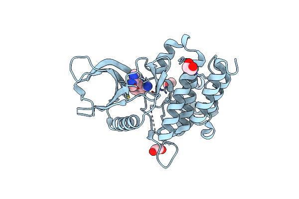 |
Organism: Homo sapiens
Method: X-RAY DIFFRACTION Resolution:1.83 Å Release Date: 2021-05-05 Classification: TRANSFERASE Ligands: EDO, RXT |
 |
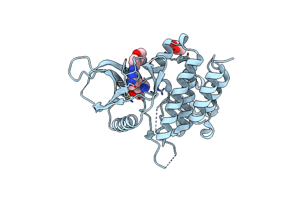
Organism: Homo sapiens
Method: X-RAY DIFFRACTION Resolution:1.74 Å Release Date: 2021-05-05 Classification: TRANSFERASE Ligands: EDO, 3JW |
 |
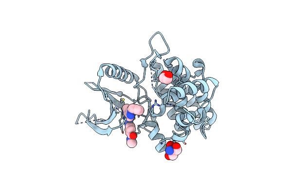
Organism: Homo sapiens
Method: X-RAY DIFFRACTION Resolution:2.50 Å Release Date: 2021-05-05 Classification: TRANSFERASE Ligands: U8P, GOL |
 |
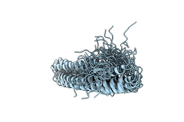
Organism: Homo sapiens
Method: X-RAY DIFFRACTION Resolution:1.80 Å Release Date: 2021-05-05 Classification: TRANSFERASE Ligands: U8J, TRS, EDO |
 |
 |
Structure Of Alpha-Galactosidase From Lactobacillus Acidophilus Ncfm, Ptcl4 Derivative
Organism: Lactobacillus acidophilus ncfm
Method: X-RAY DIFFRACTION Resolution:2.50 Å Release Date: 2011-08-10 Classification: HYDROLASE Ligands: PT, GOL |
 |
Structure Of Alpha-Galactosidase From Lactobacillus Acidophilus Ncfm With Tris
Organism: Lactobacillus acidophilus ncfm
Method: X-RAY DIFFRACTION Resolution:2.30 Å Release Date: 2011-08-10 Classification: HYDROLASE Ligands: TRS, GOL |
 |
Structure Of Alpha-Galactosidase From Lactobacillus Acidophilus Ncfm With Galactose
Organism: Lactobacillus acidophilus ncfm
Method: X-RAY DIFFRACTION Resolution:1.58 Å Release Date: 2011-08-10 Classification: HYDROLASE Ligands: GLA, IMD, GOL |
 |
The Crystal Structure Of G-Type Lysozyme From Atlantic Cod (Gadus Morhua L.) In Complex With Nag Oligomers Sheds New Light On Substrate Binding And The Catalytic Mechanism. Native Structure To 1.9
Organism: Gadus morhua
Method: X-RAY DIFFRACTION Resolution:1.90 Å Release Date: 2009-10-20 Classification: HYDROLASE Ligands: CO |
 |
The Crystal Structure Of G-Type Lysozyme From Atlantic Cod (Gadus Morhua L.) In Complex With Nag Oligomers Sheds New Light On Substrate Binding And The Catalytic Mechanism. Structure With Nag To 1.7
Organism: Gadus morhua
Method: X-RAY DIFFRACTION Resolution:1.70 Å Release Date: 2009-10-20 Classification: HYDROLASE |
 |
The 1.4 A Crystal Structure Of The Large And Cold-Active Vibrio Sp. Alkaline Phosphatase
Organism: Vibrio sp. g15-21
Method: X-RAY DIFFRACTION Resolution:1.40 Å Release Date: 2009-06-16 Classification: HYDROLASE Ligands: ZN, MG, SO4, EDO |
 |
The Refined Crystal Structure Of Lysozyme From The Rainbow Trout (Oncorhynchus Mykiss)
Organism: Oncorhynchus mykiss
Method: X-RAY DIFFRACTION Resolution:1.80 Å Release Date: 1995-02-07 Classification: HYDROLASE (O-GLYCOSYL) |