
Search Count: 16
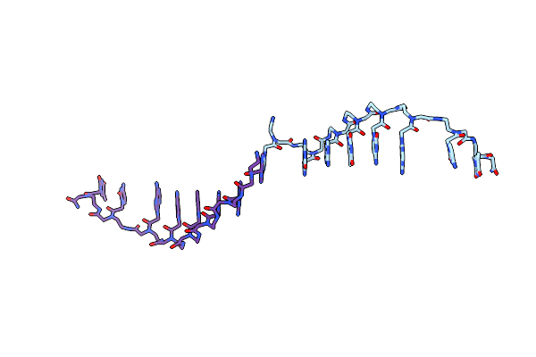 |
Crystal Structure Of A Partly Self-Complementary Peptide Nucleic Acid (Pna) Oligomer Showing A Duplex-Triplex Network
Method: X-RAY DIFFRACTION
Resolution:2.60 Å Release Date: 2005-02-22 Classification: PEPTIDE NUCLEIC ACID |
 |
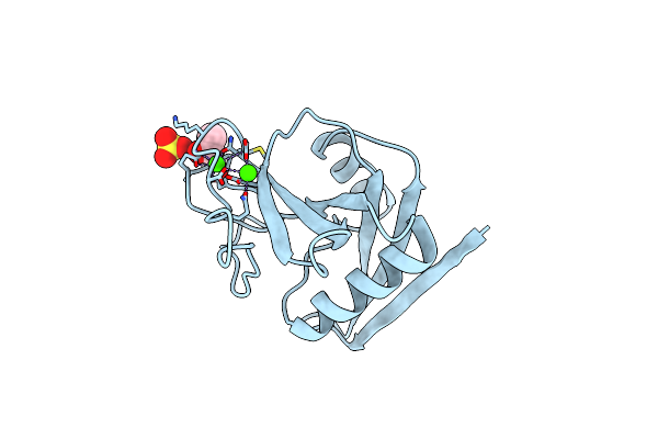
Organism: Rattus norvegicus
Method: X-RAY DIFFRACTION Resolution:2.00 Å Release Date: 2003-11-04 Classification: CELL ADHESION |
 |
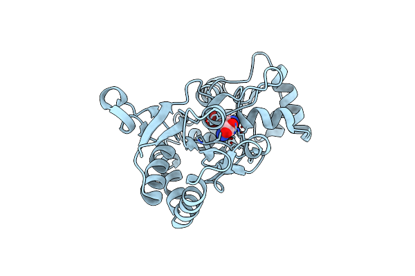
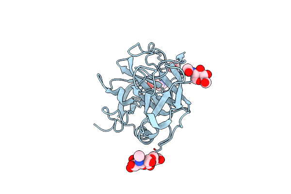
X-Ray Structure Of The Glur2 Ligand-Binding Core (S1S2J) In Complex With The Antagonist (S)-Atpo At 2.1 A Resolution.
Organism: Rattus norvegicus
Method: X-RAY DIFFRACTION Resolution:2.10 Å Release Date: 2003-03-04 Classification: MEMBRANE PROTEIN Ligands: AT1, SO4, ACT |
 |
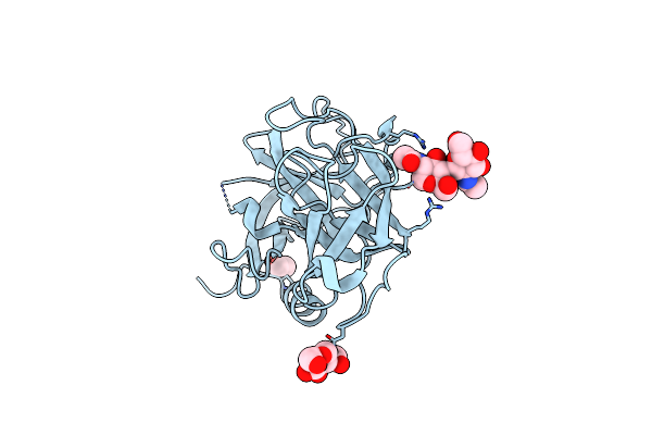
X-Ray Structure Of The Glur2 Ligand Binding Core (S1S2J) In Complex With 2-Me-Tet-Ampa At 1.85 A Resolution.
Organism: Rattus norvegicus
Method: X-RAY DIFFRACTION Resolution:1.85 Å Release Date: 2002-09-18 Classification: MEMBRANE PROTEIN Ligands: ZN, BN1 |
 |
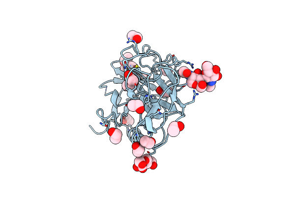
X-Ray Structure Of The Glur2 Ligand Binding Core (S1S2J) In Complex With Br-Hibo At 1.65 A Resolution
Organism: Rattus norvegicus
Method: X-RAY DIFFRACTION Resolution:1.65 Å Release Date: 2002-09-18 Classification: MEMBRANE PROTEIN Ligands: BRH |
 |
X-Ray Structure Of The Glur2 Ligand Binding Core (S1S2J-Y702F) In Complex With Br-Hibo At 1.73 A Resolution
Organism: Rattus norvegicus
Method: X-RAY DIFFRACTION Resolution:1.73 Å Release Date: 2002-09-18 Classification: MEMBRANE PROTEIN Ligands: SO4, BRH |
 |
X-Ray Structure Of The Glur2 Ligand Binding Core (S1S2J) In Complex With Acpa At 1.46 A Resolution
Organism: Rattus norvegicus
Method: X-RAY DIFFRACTION Resolution:1.46 Å Release Date: 2002-09-18 Classification: MEMBRANE PROTEIN Ligands: ZN, AM1, ACT |
 |
X-Ray Structure Of The Glur2 Ligand Binding Core (S1S2J-Y702F) In Complex With Acpa At 1.95 A Resolution
Organism: Rattus norvegicus
Method: X-RAY DIFFRACTION Resolution:1.95 Å Release Date: 2002-09-18 Classification: MEMBRANE PROTEIN Ligands: ZN, AM1, ACT |
 |
Organism: Homo sapiens
Method: X-RAY DIFFRACTION Resolution:2.50 Å Release Date: 2001-09-28 Classification: ANTIMICROBIAL PROTEIN Ligands: NAG, EOH |
 |
Organism: Homo sapiens
Method: X-RAY DIFFRACTION Resolution:1.89 Å Release Date: 2001-09-28 Classification: ANTIMICROBIAL PROTEIN Ligands: NAG, CL, EOH |
 |
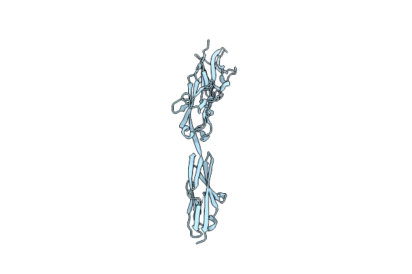
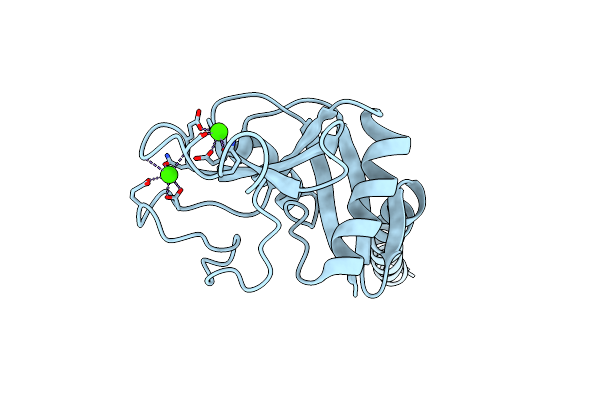
Crystal Structure Of The Two N-Terminal Immunoglobulin Domains Of The Neural Cell Adhesion Molecule (Ncam)
Organism: Rattus norvegicus
Method: X-RAY DIFFRACTION Resolution:1.85 Å Release Date: 2000-10-09 Classification: CELL ADHESION Ligands: CA |
 |
Organism: Homo sapiens
Method: X-RAY DIFFRACTION Resolution:1.12 Å Release Date: 1999-03-23 Classification: SERINE PROTEASE HOMOLOG Ligands: NAG, CL, EOH |
 |
Organism: Homo sapiens
Method: X-RAY DIFFRACTION Resolution:2.00 Å Release Date: 1998-05-06 Classification: LECTIN Ligands: CA, SO4, EOH |
 |
Organism: Homo sapiens
Method: X-RAY DIFFRACTION Resolution:2.30 Å Release Date: 1998-03-11 Classification: SERINE PROTEASE HOMOLOG Ligands: NAG |
 |
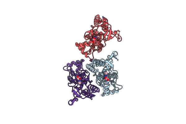
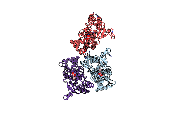
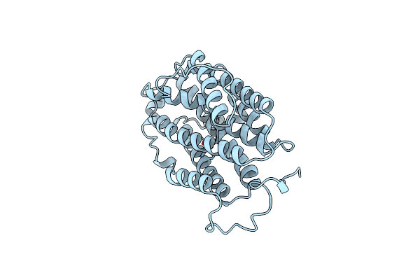
Human Tetranectin, A Trimeric Plasminogen Binding Protein With An Alpha-Helical Coiled Coil
Organism: Homo sapiens
Method: X-RAY DIFFRACTION Resolution:2.80 Å Release Date: 1997-12-03 Classification: LECTIN Ligands: CA |
 |
Organism: Mus musculus
Method: X-RAY DIFFRACTION Resolution:2.30 Å Release Date: 1997-01-11 Classification: OXIDOREDUCTASE Ligands: FE |