
Search Count: 15
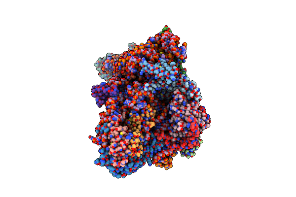 |
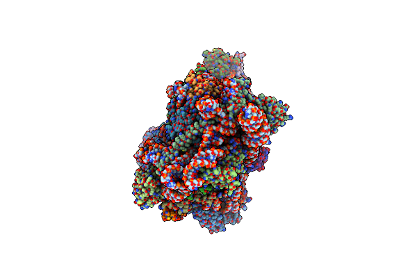
Sars-Cov-2 Nsp1 Bound To The Rhinolophus Lepidus 40S Ribosomal Subunit (Local Refinement Of The 40S Body)
Organism: Severe acute respiratory syndrome coronavirus 2, Homo sapiens, Rhinolophus lepidus
Method: ELECTRON MICROSCOPY Release Date: 2025-06-11 Classification: RIBOSOME Ligands: MG, K, ZN |
 |
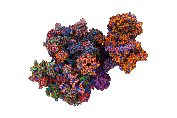
Sars-Cov-2 Nsp1 Bound To The Rhinolophus Lepidus 40S Ribosome (Local Refinement Of The 40S Head)
Organism: Rhinolophus lepidus
Method: ELECTRON MICROSCOPY Release Date: 2025-06-11 Classification: RIBOSOME Ligands: MG, ZN, K |
 |
Organism: Homo sapiens
Method: X-RAY DIFFRACTION Resolution:2.38 Å Release Date: 2024-11-27 Classification: HYDROLASE Ligands: NAG, A1IJV, ZN |
 |
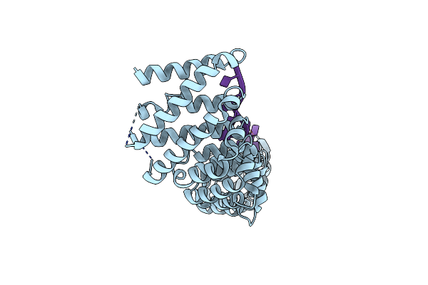
Organism: Homo sapiens
Method: X-RAY DIFFRACTION Resolution:1.99 Å Release Date: 2024-11-27 Classification: HYDROLASE Ligands: NAG, A1IJO, ZN |
 |
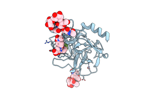
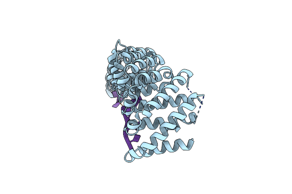
Organism: Homo sapiens
Method: X-RAY DIFFRACTION Resolution:2.20 Å Release Date: 2024-11-27 Classification: HYDROLASE Ligands: NAG, A1IJ3, ZN |
 |
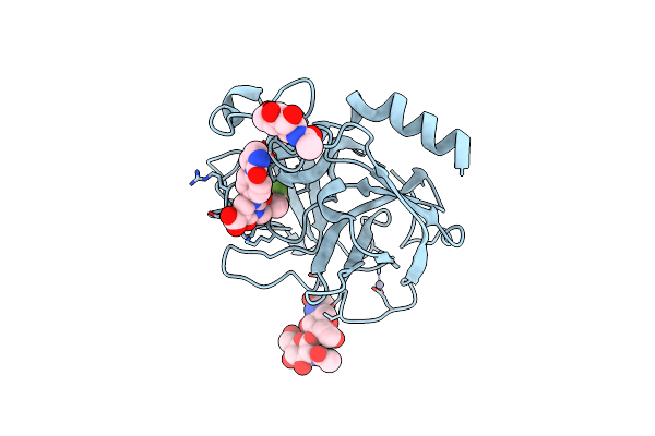
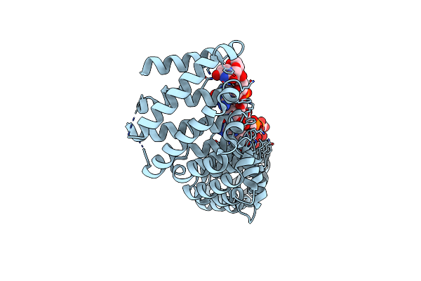
Organism: Homo sapiens
Method: X-RAY DIFFRACTION Resolution:1.79 Å Release Date: 2024-11-27 Classification: HYDROLASE Ligands: A1IJ0, ZN |
 |
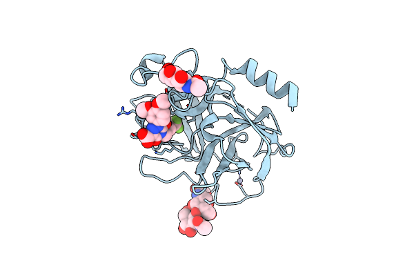
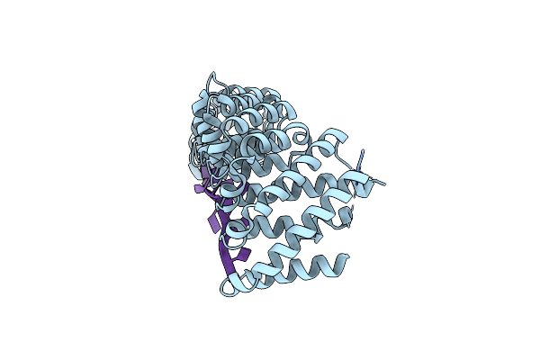
Crystal Structure Of Human Chymase In Complex With Fulacimstat (Compound86)
Organism: Homo sapiens
Method: X-RAY DIFFRACTION Resolution:1.80 Å Release Date: 2024-11-27 Classification: HYDROLASE Ligands: A1IJ2, ZN |
 |
Organism: Homo sapiens, Bat hp-betacoronavirus/zhejiang2013
Method: ELECTRON MICROSCOPY Release Date: 2023-10-18 Classification: TRANSLATION Ligands: UNX, MG, ZN |
 |
Organism: Middle east respiratory syndrome-related coronavirus, Homo sapiens
Method: ELECTRON MICROSCOPY Release Date: 2023-10-18 Classification: TRANSLATION Ligands: ZN, GTP, MG, MET, UNX |
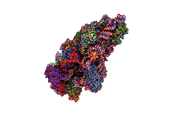 |
Cryoem Structure Of The Human 40S Small Ribosomal Subunit In Complex With Translation Initiation Factors Eif1A And Eif5B.
Organism: Homo sapiens
Method: ELECTRON MICROSCOPY Resolution:3.20 Å Release Date: 2022-04-27 Classification: RIBOSOME Ligands: GNP, 5GP, ZN |
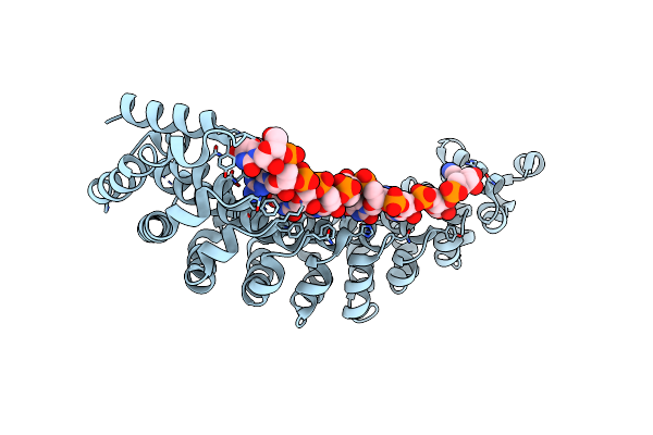 |
Crystal Structure Of The Rna-Binding Domain Of Yeast Puf5P Bound To Smx2 Rna
Organism: Saccharomyces cerevisiae (strain atcc 204508 / s288c), Synthetic construct
Method: X-RAY DIFFRACTION Resolution:2.71 Å Release Date: 2015-09-23 Classification: rna binding protein/rna |
 |
Crystal Structure Of The Rna-Binding Domain Of Yeast Puf5P Bound To Mfa2 Rna
Organism: Saccharomyces cerevisiae (strain atcc 204508 / s288c), Synthetic construct
Method: X-RAY DIFFRACTION Resolution:2.15 Å Release Date: 2015-09-23 Classification: rna binding protein/rna |
 |
Crystal Structure Of The Rna-Binding Domain Of Yeast Puf5P Bound To Amn1 Rna
Organism: Saccharomyces cerevisiae (strain atcc 204508 / s288c), Synthetic construct
Method: X-RAY DIFFRACTION Resolution:2.80 Å Release Date: 2015-09-23 Classification: rna binding protein/rna |
 |
Crystal Structure Of The Rna-Binding Domain Of Yeast Puf5P Bound To Aat2 Rna
Organism: Saccharomyces cerevisiae (strain atcc 204508 / s288c), Synthetic construct
Method: X-RAY DIFFRACTION Resolution:2.50 Å Release Date: 2015-09-23 Classification: rna binding protein/rna |
 |
Crystal Structure Of The Rna-Binding Domain Of Yeast Puf5P Bound To Smx2 Rna
Organism: Saccharomyces cerevisiae (strain atcc 204508 / s288c), Synthetic construct
Method: X-RAY DIFFRACTION Resolution:2.35 Å Release Date: 2015-09-23 Classification: rna binding protein/rna |