
Search Count: 21
 |
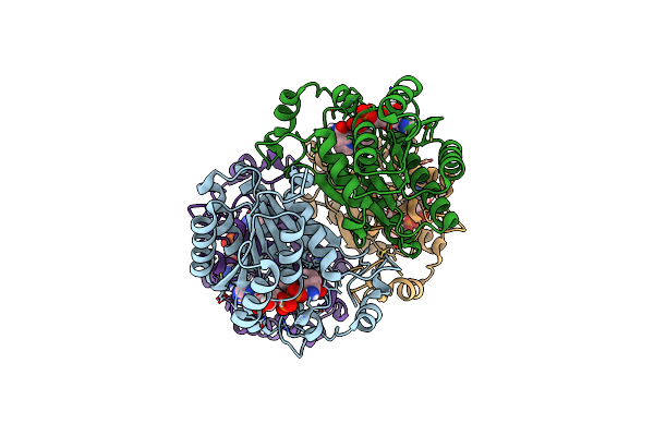
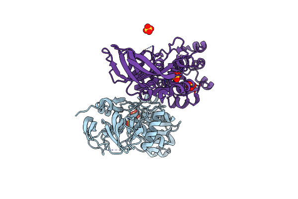
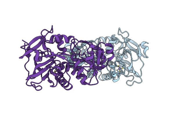
Proline Activating Adenylation Domain Of Gramicidin S Synthetase 2 - Grsb1-Acore
Organism: Aneurinibacillus migulanus
Method: X-RAY DIFFRACTION Resolution:2.60 Å Release Date: 2023-07-05 Classification: BIOSYNTHETIC PROTEIN |
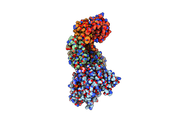 |
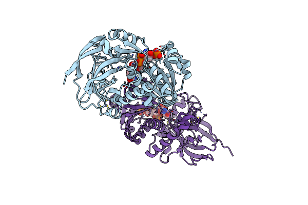
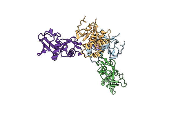
Cryo-Em Structure Of Human Apobec3G/Hiv-1 Vif/Cbfbeta/Elob/Eloc Monomeric Complex
Organism: Homo sapiens, Human immunodeficiency virus 1, Spodoptera frugiperda
Method: ELECTRON MICROSCOPY Release Date: 2023-02-15 Classification: VIRAL PROTEIN/IMMUNE SYSTEM/RNA Ligands: ZN |
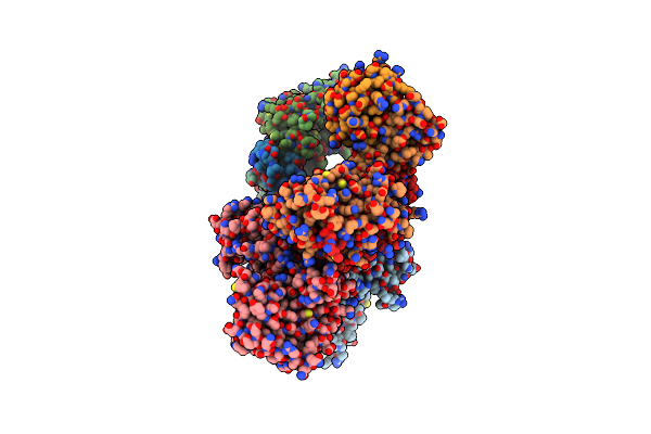 |
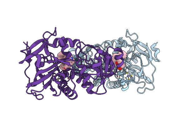
Cryo-Em Structure Of Human Apobec3G/Hiv-1 Vif/Cbfbeta/Elob/Eloc Dimeric Complex In State 1
Organism: Homo sapiens, Human immunodeficiency virus 1, Spodoptera frugiperda
Method: ELECTRON MICROSCOPY Release Date: 2023-02-15 Classification: VIRAL PROTEIN/IMMUNE SYSTEM/RNA Ligands: ZN |
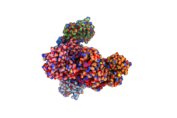 |
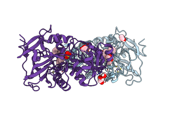
Cryo-Em Structure Of Human Apobec3G/Hiv-1 Vif/Cbfbeta/Elob/Eloc Dimeric Complex In State 2
Organism: Homo sapiens, Human immunodeficiency virus 1, Spodoptera frugiperda
Method: ELECTRON MICROSCOPY Release Date: 2023-02-15 Classification: VIRAL PROTEIN/IMMUNE SYSTEM/RNA Ligands: ZN |
 |
Organism: Nepeta sibirica
Method: X-RAY DIFFRACTION Resolution:1.85 Å Release Date: 2022-12-28 Classification: BIOSYNTHETIC PROTEIN Ligands: NAD |
 |
Organism: Catharanthus roseus
Method: X-RAY DIFFRACTION Resolution:2.45 Å Release Date: 2022-11-02 Classification: CYTOSOLIC PROTEIN Ligands: SO4 |
 |
Geissoschizine Synthase From Catharanthus Roseus - Binary Complex With Nadp+
Organism: Catharanthus roseus
Method: X-RAY DIFFRACTION Resolution:2.00 Å Release Date: 2022-10-19 Classification: OXIDOREDUCTASE Ligands: NAP, ZN |
 |
Organism: Tabernanthe iboga
Method: X-RAY DIFFRACTION Resolution:1.88 Å Release Date: 2022-10-19 Classification: CYTOSOLIC PROTEIN Ligands: ORZ, ZN |
 |
Dihydroprecondylocarpine Acetate Synthase 2 From Tabernanthe Iboga - Stemmadenine Acetate Bound Structure
Organism: Tabernanthe iboga
Method: X-RAY DIFFRACTION Resolution:2.24 Å Release Date: 2022-10-19 Classification: CYTOSOLIC PROTEIN Ligands: OSO, EDO, SO4, ZN |
 |
Organism: Tabernanthe iboga
Method: X-RAY DIFFRACTION Resolution:2.42 Å Release Date: 2022-10-19 Classification: CYTOSOLIC PROTEIN Ligands: ZN |
 |
Crystal Structure Of Recombinant Human Acetylcholinesterase In Complex With Gd And Hi-6
Organism: Homo sapiens
Method: X-RAY DIFFRACTION Resolution:2.19 Å Release Date: 2021-03-31 Classification: HYDROLASE Ligands: EDO, GD8, HI6, NAG, NO3, PE8 |
 |
Crystal Structure Of Recombinant Human Acetylcholinesterase Inhibited By Gb
Organism: Homo sapiens
Method: X-RAY DIFFRACTION Resolution:2.25 Å Release Date: 2021-02-24 Classification: HYDROLASE Ligands: UCJ, 7PE, NAG |
 |
Crystal Structure Of Recombinant Human Acetylcholinesterase Inhibited By Ga
Organism: Homo sapiens
Method: X-RAY DIFFRACTION Resolution:2.63 Å Release Date: 2021-02-17 Classification: HYDROLASE Ligands: NAG, ELT |
 |
Crystal Structure Of Recombinant Human Acetylcholinesterase In Complex With Ga And Hi-6
Organism: Homo sapiens
Method: X-RAY DIFFRACTION Resolution:2.46 Å Release Date: 2021-02-17 Classification: HYDROLASE Ligands: NAG, ELT, HI6 |
 |
Crystal Structure Of Recombinant Human Acetylcholinesterase In Complex With Gb And Hi-6
Organism: Homo sapiens
Method: X-RAY DIFFRACTION Resolution:2.37 Å Release Date: 2021-02-17 Classification: HYDROLASE Ligands: NAG, 7PE, UCJ, HI6 |
 |
Crystal Structure Of Recombinant Human Acetylcholinesterase Inhibited By Gd
Organism: Homo sapiens
Method: X-RAY DIFFRACTION Resolution:2.60 Å Release Date: 2021-02-17 Classification: HYDROLASE Ligands: NAG, UCY, 7PE |
 |
Crystal Structure Of Recombinant Human Acetylcholinesterase Inhibited By Gf
Organism: Homo sapiens
Method: X-RAY DIFFRACTION Resolution:2.31 Å Release Date: 2021-02-17 Classification: HYDROLASE Ligands: NAG, WW2, 7PE |
 |
Crystal Structure Of Recombinant Human Acetylcholinesterase Inhibited By Gp
Organism: Homo sapiens
Method: X-RAY DIFFRACTION Resolution:2.29 Å Release Date: 2021-02-17 Classification: HYDROLASE Ligands: NAG, 7PE, UCY |
 |
Structure Of The Kupe Virus Otu Bound To The C-Terminal Domain Of Sheep Isg15
Organism: Kupe virus, Ovis aries
Method: X-RAY DIFFRACTION Resolution:2.06 Å Release Date: 2020-01-08 Classification: HYDROLASE Ligands: AYE |
 |
Organism: Ganjam virus, Ovis aries
Method: X-RAY DIFFRACTION Resolution:3.17 Å Release Date: 2020-01-08 Classification: HYDROLASE Ligands: AYE |