
Search Count: 6
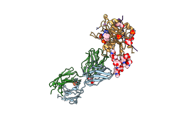 |
Organism: Homo sapiens
Method: X-RAY DIFFRACTION Resolution:1.90 Å Release Date: 2015-10-28 Classification: IMMUNE SYSTEM/HYDROLASE Ligands: PEG, PO4, CIT, NA, 0G6 |
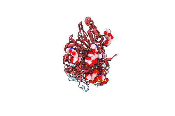 |
Organism: Homo sapiens
Method: X-RAY DIFFRACTION Resolution:1.70 Å Release Date: 2010-02-02 Classification: HYDROLASE, HYDROLASE INHIBITOR Ligands: CA, MPD |
 |
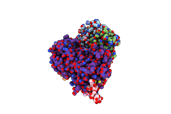
Intermediate Structure Of Antithrombin Bound To The Natural Pentasaccharide
Organism: Homo sapiens
Method: X-RAY DIFFRACTION Resolution:3.00 Å Release Date: 2008-10-21 Classification: HYDROLASE inhibitor Ligands: NAG |
 |
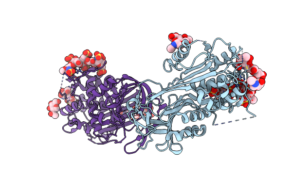
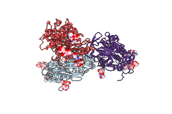
2.1 Angstrom Structure Of A Nonproductive Complex Between Antithrombin, Synthetic Heparin Mimetic Sr123781 And Two S195A Thrombin Molecules
Organism: Homo sapiens
Method: X-RAY DIFFRACTION Resolution:2.10 Å Release Date: 2006-09-19 Classification: BLOOD CLOTTING Ligands: GOL, SO4, NAG |
 |
Crystal Structure Of Antithrombin Variant S137A/V317C/T401C With Plasma Latent Antithrombin
Organism: Homo sapiens
Method: X-RAY DIFFRACTION Resolution:2.70 Å Release Date: 2005-11-01 Classification: BLOOD CLOTTING Ligands: NAG, GOL |
 |
Organism: Homo sapiens
Method: X-RAY DIFFRACTION Resolution:2.75 Å Release Date: 2005-10-04 Classification: BLOOD CLOTTING Ligands: IOD, GOL |