
Search Count: 494
 |
Organism: Human gammaherpesvirus 4
Method: ELECTRON MICROSCOPY Release Date: 2025-12-31 Classification: VIRAL PROTEIN |
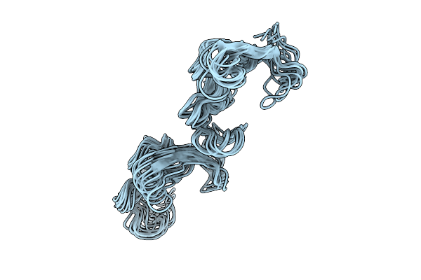 |
Organism: Gallus gallus
Method: SOLUTION NMR Release Date: 2025-12-31 Classification: STRUCTURAL PROTEIN |
 |
Organism: Homo sapiens, Human gammaherpesvirus 4
Method: ELECTRON MICROSCOPY Release Date: 2025-12-31 Classification: VIRAL PROTEIN Ligands: NAG |
 |
Organism: Monkeypox virus, Homo sapiens
Method: ELECTRON MICROSCOPY Release Date: 2025-12-24 Classification: VIRAL PROTEIN |
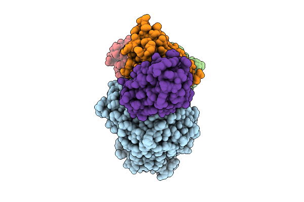 |
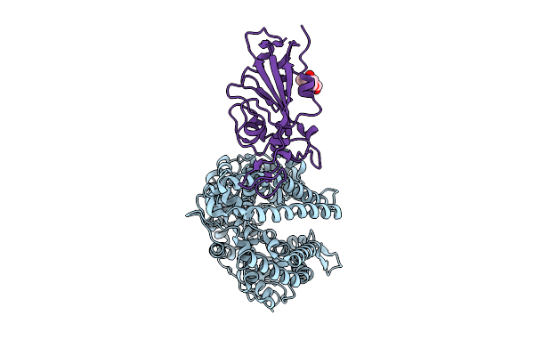
Globular Domain Of Monkeypox Virus Opg153 (A28) In Complex With Antibodies 08E11 And 12I12
Organism: Monkeypox virus, Homo sapiens
Method: ELECTRON MICROSCOPY Release Date: 2025-12-24 Classification: VIRAL PROTEIN |
 |
Organism: Gallus gallus
Method: X-RAY DIFFRACTION Release Date: 2025-11-26 Classification: MEMBRANE PROTEIN Ligands: NAG, EDO |
 |
Organism: Ovis aries, Bat coronavirus ratg13
Method: X-RAY DIFFRACTION Release Date: 2025-11-26 Classification: VIRAL PROTEIN/HYDROLASE Ligands: NAG |
 |
Organism: Bos taurus, Bat coronavirus ratg13
Method: X-RAY DIFFRACTION Release Date: 2025-11-26 Classification: VIRAL PROTEIN/HYDROLASE Ligands: NAG |
 |
Organism: Plasmodium falciparum 3d7
Method: ELECTRON MICROSCOPY Release Date: 2025-11-12 Classification: CYTOSOLIC PROTEIN Ligands: A1B74 |
 |
Organism: Plasmodium falciparum 3d7
Method: ELECTRON MICROSCOPY Release Date: 2025-11-12 Classification: CYTOSOLIC PROTEIN Ligands: A1B73 |
 |
Organism: Danio rerio
Method: ELECTRON MICROSCOPY Release Date: 2025-11-05 Classification: MEMBRANE PROTEIN Ligands: Y01, A1BKU, CLR |
 |
Organism: Danio rerio
Method: ELECTRON MICROSCOPY Release Date: 2025-11-05 Classification: MEMBRANE PROTEIN Ligands: Y01, QNJ |
 |
Organism: Danio rerio
Method: ELECTRON MICROSCOPY Release Date: 2025-11-05 Classification: MEMBRANE PROTEIN Ligands: Y01, CLR, A1BKT |
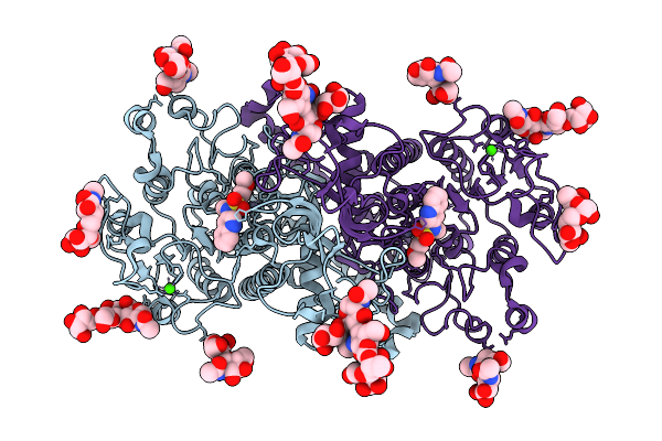 |
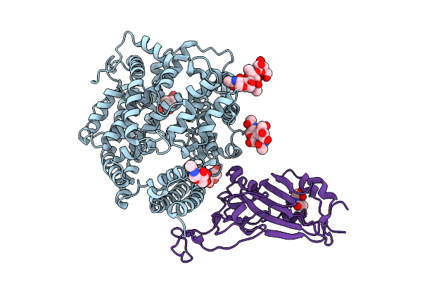
The Cryo-Em Structure Of Human Tissue Nonspecific Alkaline Phosphatase (Htnap) In Complex With Mls-0038949
Organism: Homo sapiens
Method: ELECTRON MICROSCOPY Release Date: 2025-11-05 Classification: HYDROLASE Ligands: MG, ZN, CA, A1JNY |
 |
Structure Of The Respiratory Syncytial Virus Fusion Protein Bound To Human Antibodies Rsv_2245 And Rsv_3301
Organism: Respiratory syncytial virus, Homo sapiens
Method: ELECTRON MICROSCOPY Release Date: 2025-10-29 Classification: VIRAL PROTEIN/IMMUNE SYSTEM |
 |
Crystal Structure Of Ratg13 Receptor-Binding Domain Complexed With Squirrel Ace2
Organism: Petaurus norfolcensis, Bat coronavirus ratg13
Method: X-RAY DIFFRACTION Release Date: 2025-08-06 Classification: VIRAL PROTEIN Ligands: CL, NAG, ZN |
 |
Crystal Structure Of Pcov-Gd Receptor-Binding Domain Complexed With Squirrel Ace2
Organism: Pangolin coronavirus, Petaurus norfolcensis
Method: X-RAY DIFFRACTION Release Date: 2025-08-06 Classification: VIRAL PROTEIN Ligands: NAG |
 |
Crystal Structure Of Pcov-Gx Receptor-Binding Domain Complexed With Squirrel Ace2
Organism: Petaurus norfolcensis, Pangolin coronavirus
Method: X-RAY DIFFRACTION Release Date: 2025-08-06 Classification: VIRAL PROTEIN Ligands: NAG |
 |
Crystal Structure Of Sars-Cov-2 Receptor-Binding Domain Complexed With Squirrel Ace2
Organism: Petaurus norfolcensis, Severe acute respiratory syndrome coronavirus 2
Method: X-RAY DIFFRACTION Release Date: 2025-08-06 Classification: VIRAL PROTEIN Ligands: NAG, ZN |
 |
Crystal Structure Of Sars-Cov-2 Jn.1 Variant Rbd Complexed With Squirrel Ace2
Organism: Petaurus norfolcensis, Severe acute respiratory syndrome coronavirus 2
Method: X-RAY DIFFRACTION Release Date: 2025-08-06 Classification: VIRAL PROTEIN/HYDROLASE Ligands: NAG, ZN |