
Search Count: 12
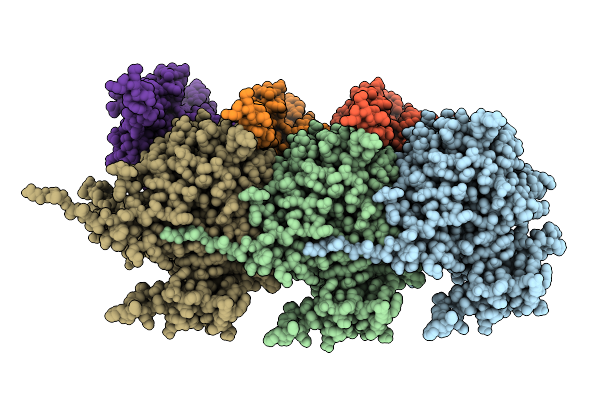 |
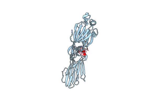
Organism: Porphyromonas gingivalis atcc 33277
Method: ELECTRON MICROSCOPY Resolution:3.20 Å Release Date: 2025-08-06 Classification: MEMBRANE PROTEIN |
 |
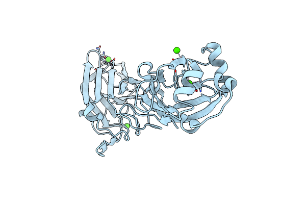
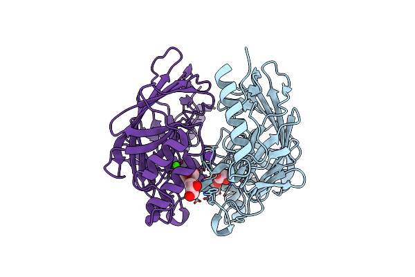
Crystal Structure Of Dimeric Porphyromonas Gingivalis Porx, A Type 9 Secretion System Response Regulator.
Organism: Porphyromonas gingivalis (strain atcc baa-308 / w83)
Method: X-RAY DIFFRACTION Resolution:2.41 Å Release Date: 2022-12-14 Classification: SIGNALING PROTEIN Ligands: BEF, MG, ZN, GOL, FMT |
 |
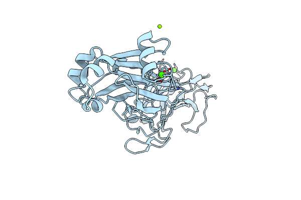
1.9 Angstrom Crystal Structure Of Dimeric Porx, Co-Crystallized In The Presence Of Zinc
Organism: Porphyromonas gingivalis (strain atcc baa-308 / w83)
Method: X-RAY DIFFRACTION Resolution:1.91 Å Release Date: 2022-12-14 Classification: SIGNALING PROTEIN Ligands: MG, BEF, ZN, GOL, FMT, CL |
 |
Organism: Porphyromonas gingivalis w83, Synthetic construct
Method: X-RAY DIFFRACTION Resolution:2.40 Å Release Date: 2022-12-14 Classification: SIGNALING PROTEIN Ligands: MG, BEF, ZN, GOL, FMT |
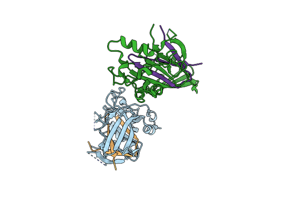 |
Organism: Streptococcus gordonii (strain challis / atcc 35105 / bcrc 15272 / ch1 / dl1 / v288)
Method: X-RAY DIFFRACTION Resolution:2.66 Å Release Date: 2016-12-14 Classification: CELL ADHESION |
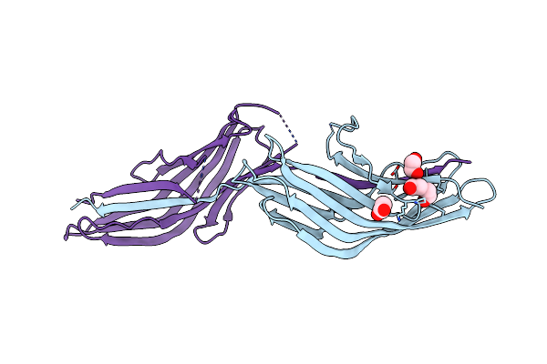 |
Organism: Streptococcus agalactiae serotype iii (strain nem316)
Method: X-RAY DIFFRACTION Resolution:2.41 Å Release Date: 2016-06-22 Classification: CELL ADHESION Ligands: ACT, PEG, EDO |
 |
Organism: Streptococcus agalactiae serotype iii (strain nem316)
Method: X-RAY DIFFRACTION Resolution:1.89 Å Release Date: 2016-06-22 Classification: CELL ADHESION |
 |
Organism: Streptococcus agalactiae serotype iii (strain nem316)
Method: X-RAY DIFFRACTION Resolution:2.19 Å Release Date: 2016-06-22 Classification: CELL ADHESION Ligands: EDO, PEG |
 |
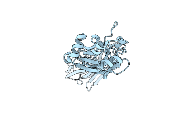
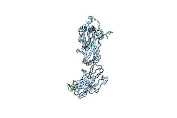
Crystal Structure Of The C-Terminal Domain Of Streptococcus Gordonii Surface Protein Sspb
Organism: Streptococcus gordonii
Method: X-RAY DIFFRACTION Resolution:1.50 Å Release Date: 2010-02-16 Classification: CELL ADHESION Ligands: CA |
 |
Crystal Structure Of The C-Terminal Domain Of Streptococcus Gordonii Surface Protein Sspb
Organism: Streptococcus gordonii
Method: X-RAY DIFFRACTION Resolution:1.70 Å Release Date: 2010-02-16 Classification: CELL ADHESION Ligands: CA, MG |
 |
Two Intramolecular Isopeptide Bonds Are Identified In The Crystal Structure Of The Streptococcus Gordonii Sspb C-Terminal Domain
Organism: Streptococcus gordonii
Method: X-RAY DIFFRACTION Resolution:2.08 Å Release Date: 2010-02-16 Classification: CELL ADHESION Ligands: CA |
 |
Crystal Structure Of The Variable Domain Of The Streptococcus Gordonii Surface Protein Sspb
Organism: Streptococcus gordonii
Method: X-RAY DIFFRACTION Resolution:2.30 Å Release Date: 2009-07-28 Classification: CELL ADHESION Ligands: CA, GOL, K |