
Search Count: 16
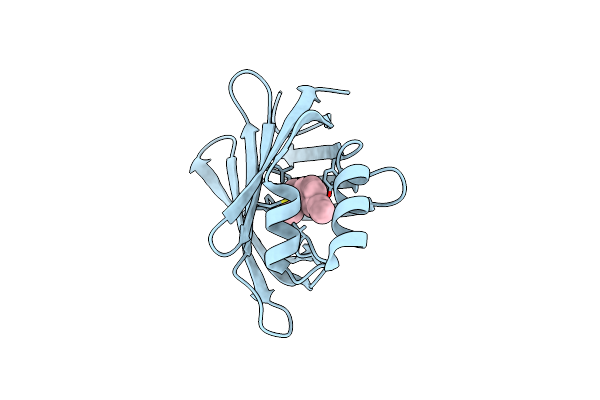 |
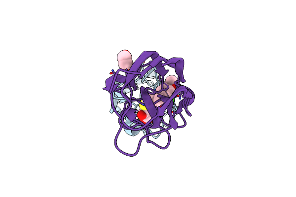
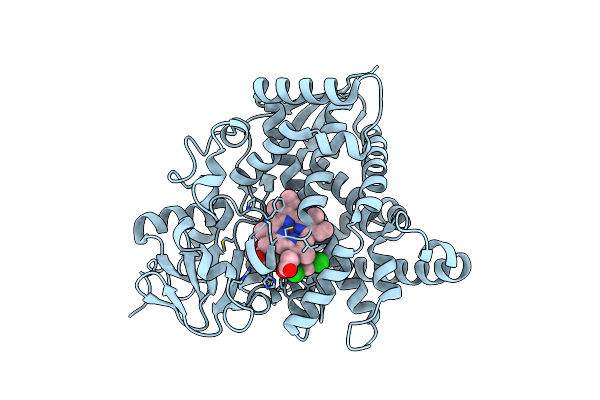
M2 Mutant (R111K:Y134F:T54V:R132Q:P39Y:R59Y) Of Human Cellular Retinoic Acid Binding Protein Ii - 1E Conjugate
Organism: Homo sapiens
Method: X-RAY DIFFRACTION Resolution:2.10 Å Release Date: 2025-03-26 Classification: TRANSPORT PROTEIN Ligands: A1IQZ |
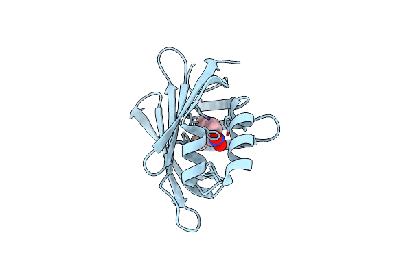 |
M2 Mutant (R111K:Y134F:T54V:R132Q:P39Y:R59Y) Of Human Cellular Retinoic Acid Binding Protein Ii - 1M Conjugate
Organism: Homo sapiens
Method: X-RAY DIFFRACTION Resolution:2.05 Å Release Date: 2025-03-26 Classification: TRANSPORT PROTEIN Ligands: A1IQY |
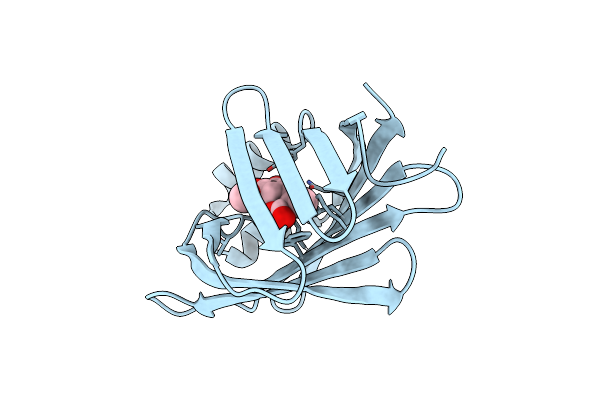 |
M2 Mutant (R111K:Y134F:T54V:R132Q:P39Y:R59Y) Of Human Cellular Retinoic Acid Binding Protein Ii - 1J Conjugate
Organism: Homo sapiens
Method: X-RAY DIFFRACTION Resolution:3.00 Å Release Date: 2025-03-26 Classification: TRANSPORT PROTEIN Ligands: A1IQ0 |
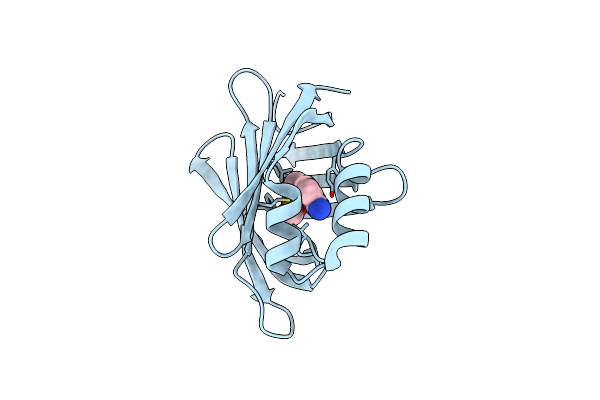 |
M2 Mutant (R111K:Y134F:T54V:R132Q:P39Y:R59Y) Of Human Cellular Retinoic Acid Binding Protein Ii - 1L Conjugate
Organism: Homo sapiens
Method: X-RAY DIFFRACTION Resolution:2.40 Å Release Date: 2025-03-26 Classification: TRANSPORT PROTEIN Ligands: A1IQ1 |
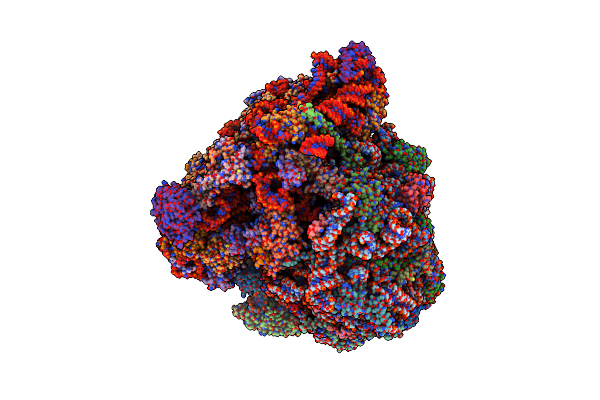 |
Cryo-Em Structure Of Leishmania Major 80S Ribosome With A/P/E-Site Trna And Mrna : Parental Strain
Organism: Leishmania major strain friedlin
Method: ELECTRON MICROSCOPY Release Date: 2024-05-15 Classification: RIBOSOME Ligands: MG, NA, K, A1H4F, ZN |
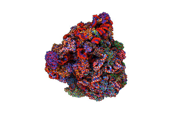 |
Cryo-Em Structure Of Leishmania Major 80S Ribosome With A/P/E-Site Trna And Mrna : Lm32Cs3H1 Sko Strain
Organism: Leishmania major strain friedlin
Method: ELECTRON MICROSCOPY Release Date: 2024-05-15 Classification: RIBOSOME Ligands: K, NA, MG, ZN |
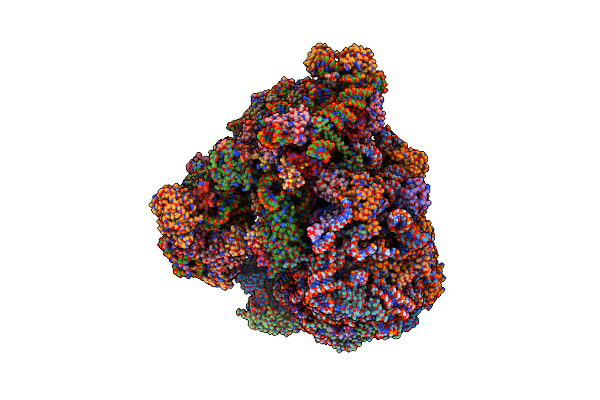 |
Organism: Leishmania major strain friedlin
Method: ELECTRON MICROSCOPY Release Date: 2024-05-08 Classification: RIBOSOME Ligands: MG, K, NA, ZN |
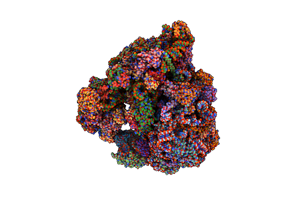 |
Organism: Leishmania major strain friedlin
Method: ELECTRON MICROSCOPY Release Date: 2023-10-11 Classification: RIBOSOME Ligands: MG, ZN, NA, K |
 |
Structure Of The Leca Lectin From Pseudomonas Aeruginosa In Complex With A Biaryl-Thiogalactoside
Organism: Pseudomonas aeruginosa pao1
Method: X-RAY DIFFRACTION Resolution:1.53 Å Release Date: 2023-01-18 Classification: SUGAR BINDING PROTEIN Ligands: CA, IEC, EDO |
 |
Structure Of The Leca Lectin From Pseudomonas Aeruginosa In Complex With A Biaryl-Thiogalactoside
Organism: Pseudomonas aeruginosa pao1
Method: X-RAY DIFFRACTION Resolution:1.75 Å Release Date: 2023-01-18 Classification: SUGAR BINDING PROTEIN Ligands: CA, IE3, EDO |
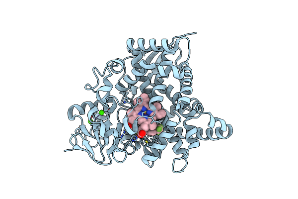 |
Naegleria Fowleri Cyp51 (Nfcyp51) Complex With (S)-1-(4-Fluorophenyl)-2-(1H-Imidazol-1-Yl)Ethyl 3-(Trifluoromethyl)Benzoate
Organism: Naegleria fowleri
Method: X-RAY DIFFRACTION Resolution:1.76 Å Release Date: 2022-07-27 Classification: OXIDOREDUCTASE Ligands: HEM, L49, CA |
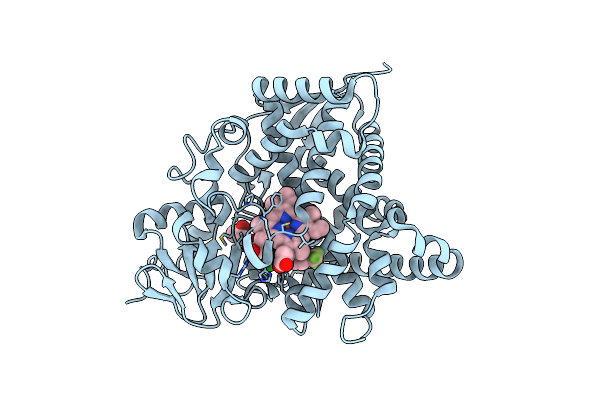 |
Naegleria Fowleri Cyp51 (Nfcyp51) Complex With (S)-1-(2,4-Dichlorophenyl)-2-(1H-Imidazol-1-Yl)Ethyl 3-(Trifluoromethyl)Benzoate
Organism: Naegleria fowleri
Method: X-RAY DIFFRACTION Resolution:2.10 Å Release Date: 2022-07-27 Classification: OXIDOREDUCTASE Ligands: HEM, 5UR, GOL |
 |
Naegleria Fowleri Cyp51(Nfcyp51) Complex With (S)-1-(4-Fluorophenyl)-2-(1H-Imidazol-1-Yl)Ethyl 3,5-Dichlorobenzoate
Organism: Naegleria fowleri
Method: X-RAY DIFFRACTION Resolution:1.81 Å Release Date: 2022-07-27 Classification: OXIDOREDUCTASE Ligands: HEM, 5TV, GOL |
 |
1,1-Diheterocyclic Ethylenes Derived From Quinaldine And Carbazole As New Tubulin Polymerization Inhibitors: Synthesis, Metabolism, And Biological Evaluation
Organism: Rattus norvegicus, Ovis aries
Method: X-RAY DIFFRACTION Resolution:2.75 Å Release Date: 2018-12-19 Classification: TRANSPORT PROTEIN Ligands: SO4, GTP, MG, GDP, FWH |
 |
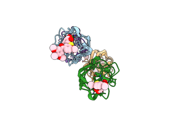
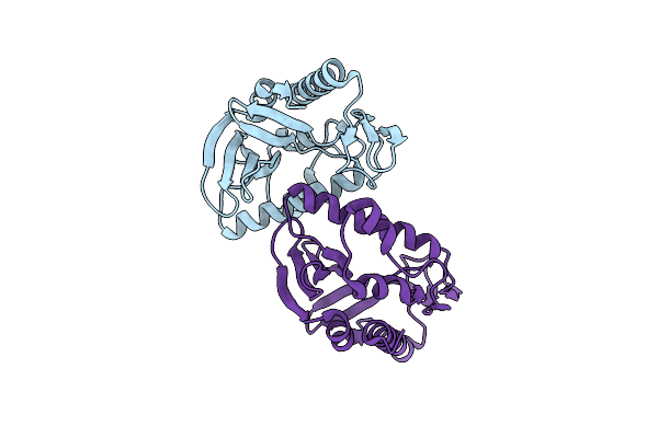
Crystal Structure Of Human Baff In Complex With Fab Fragment Of Anti-Baff Antibody Belimumab
Organism: Homo sapiens
Method: X-RAY DIFFRACTION Resolution:2.90 Å Release Date: 2018-04-04 Classification: IMMUNE SYSTEM |
 |
Organism: Escherichia coli
Method: X-RAY DIFFRACTION Resolution:2.00 Å Release Date: 2009-01-27 Classification: PROTEIN BINDING |