
Search Count: 50
 |
Organism: Mycobacterium tuberculosis, Aequorea victoria, Mycolicibacterium smegmatis mc2 155
Method: ELECTRON MICROSCOPY Release Date: 2025-10-08 Classification: MEMBRANE PROTEIN |
 |
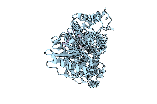
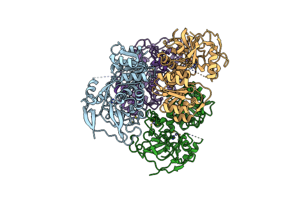
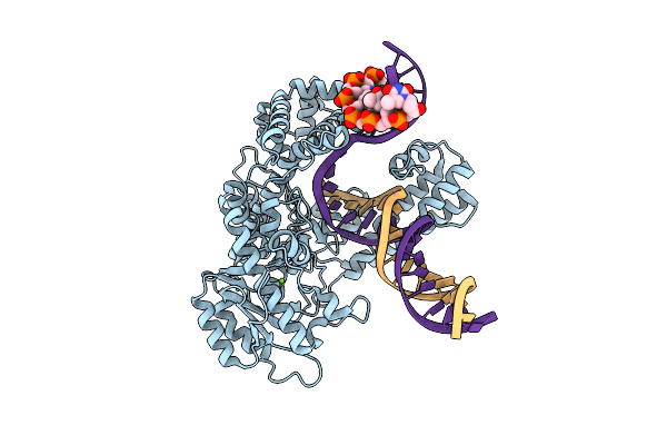
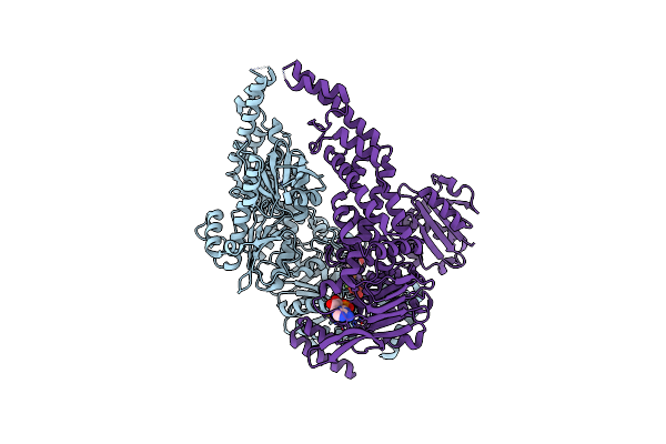
Organism: Enterococcus faecium, Escherichia coli
Method: ELECTRON MICROSCOPY Release Date: 2025-09-24 Classification: DNA BINDING PROTEIN Ligands: A1I88, ZN, MG |
 |
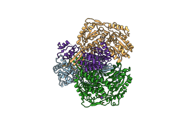
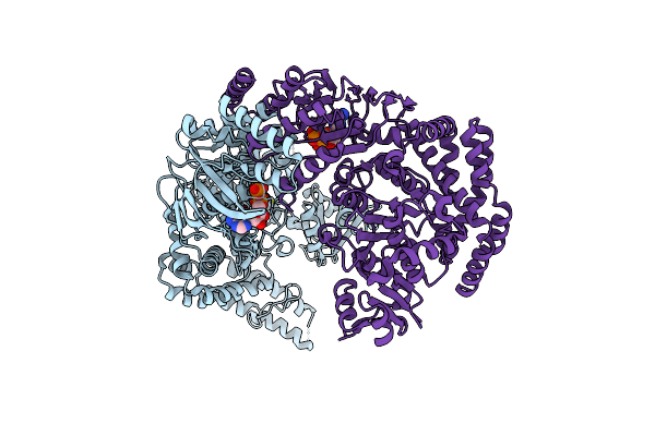
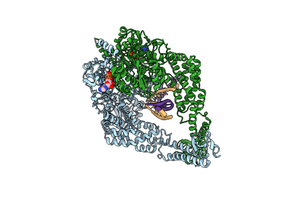
Organism: Enterococcus faecium, Escherichia coli
Method: ELECTRON MICROSCOPY Release Date: 2025-09-24 Classification: DNA BINDING PROTEIN Ligands: ZN, MG, A1I9T |
 |
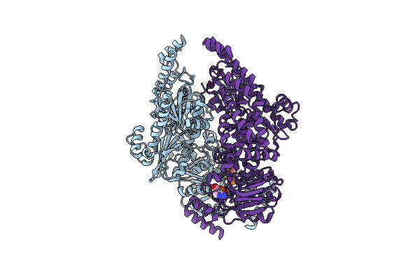
Organism: Enterococcus faecium, Escherichia coli
Method: ELECTRON MICROSCOPY Release Date: 2025-09-24 Classification: DNA BINDING PROTEIN Ligands: ZN, MG |
 |
Organism: Mycobacterium tuberculosis h37rv, Mycobacterium tuberculosis, Mycolicibacterium smegmatis mc2 155
Method: ELECTRON MICROSCOPY Release Date: 2025-08-13 Classification: MEMBRANE PROTEIN Ligands: L9Q |
 |
Organism: Mycobacterium tuberculosis, Aequorea victoria, Mycolicibacterium smegmatis mc2 155
Method: ELECTRON MICROSCOPY Release Date: 2025-08-13 Classification: MEMBRANE PROTEIN Ligands: L9Q |
 |
Organism: Paracoccus denitrificans
Method: ELECTRON MICROSCOPY Release Date: 2025-06-11 Classification: OXIDOREDUCTASE Ligands: ZN |
 |
Organism: Paracoccus denitrificans
Method: ELECTRON MICROSCOPY Release Date: 2025-06-11 Classification: OXIDOREDUCTASE Ligands: K |
 |
Organism: Mycolicibacterium smegmatis mc2 155
Method: ELECTRON MICROSCOPY Release Date: 2025-04-09 Classification: OXIDOREDUCTASE Ligands: FES, 9YF, PLM, HEM, CDL, A1IRE, HEC, MQ9, 9Y0, CU, HEA, CA, MQ7 |
 |
Organism: Escherichia coli 'bl21-gold(de3)plyss ag', Synthetic construct
Method: ELECTRON MICROSCOPY Release Date: 2023-08-09 Classification: DNA BINDING PROTEIN Ligands: MG |
 |
Organism: Escherichia coli 'bl21-gold(de3)plyss ag', Synthetic construct
Method: ELECTRON MICROSCOPY Release Date: 2023-08-09 Classification: DNA BINDING PROTEIN Ligands: MG |
 |
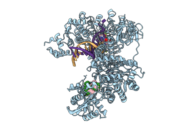
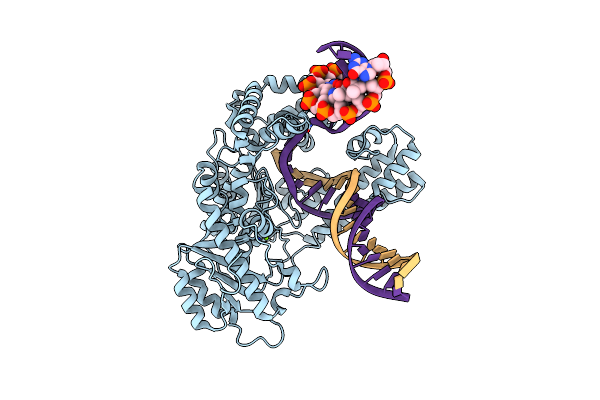
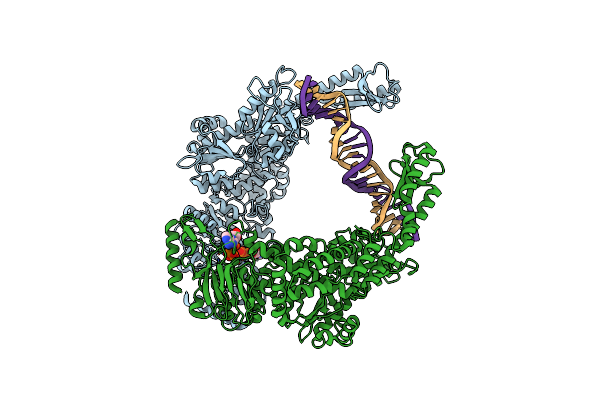
Organism: Escherichia coli (strain k12), Dna molecule
Method: ELECTRON MICROSCOPY Resolution:3.60 Å Release Date: 2022-06-29 Classification: DNA BINDING PROTEIN Ligands: ANP, MG |
 |
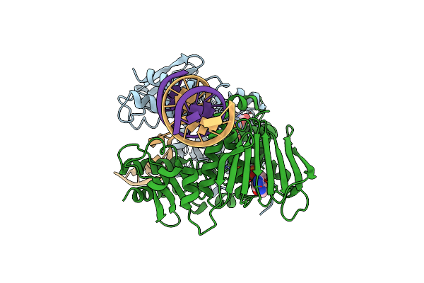
Organism: Mycobacterium tuberculosis, Synthetic construct
Method: ELECTRON MICROSCOPY Release Date: 2022-02-23 Classification: REPLICATION Ligands: ZN, 82W |
 |
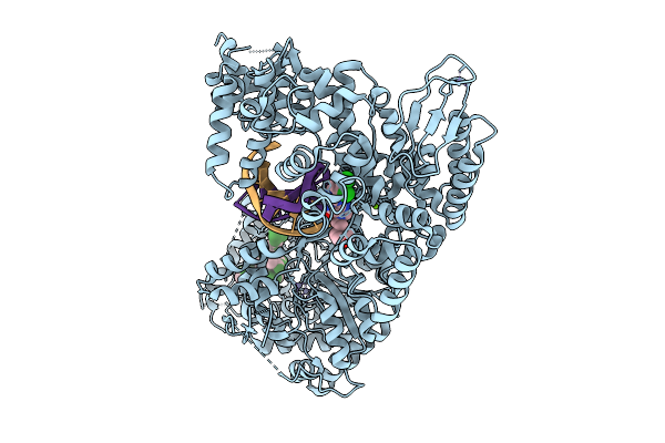
Organism: Escherichia coli (strain k12)
Method: ELECTRON MICROSCOPY Release Date: 2022-01-12 Classification: DNA BINDING PROTEIN Ligands: MG, ANP |
 |
Organism: Escherichia coli (strain k12)
Method: ELECTRON MICROSCOPY Release Date: 2022-01-12 Classification: DNA BINDING PROTEIN Ligands: MG, ADP, VO4 |
 |
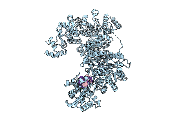
Organism: Escherichia coli
Method: ELECTRON MICROSCOPY Release Date: 2022-01-12 Classification: DNA BINDING PROTEIN Ligands: ADP |
 |
Organism: Escherichia coli
Method: ELECTRON MICROSCOPY Resolution:3.30 Å Release Date: 2022-01-12 Classification: DNA BINDING PROTEIN Ligands: MG, ATP, ADP |
 |
Organism: Escherichia coli, Synthetic construct
Method: ELECTRON MICROSCOPY Resolution:4.40 Å Release Date: 2021-03-31 Classification: DNA BINDING PROTEIN Ligands: ATP |
 |
Organism: Escherichia coli (strain k12), Synthetic construct
Method: ELECTRON MICROSCOPY Release Date: 2021-03-31 Classification: DNA BINDING PROTEIN Ligands: ADP |
 |
Organism: Escherichia coli (strain k12), Synthetic construct
Method: ELECTRON MICROSCOPY Release Date: 2021-03-31 Classification: DNA BINDING PROTEIN Ligands: ADP |