
Search Count: 136
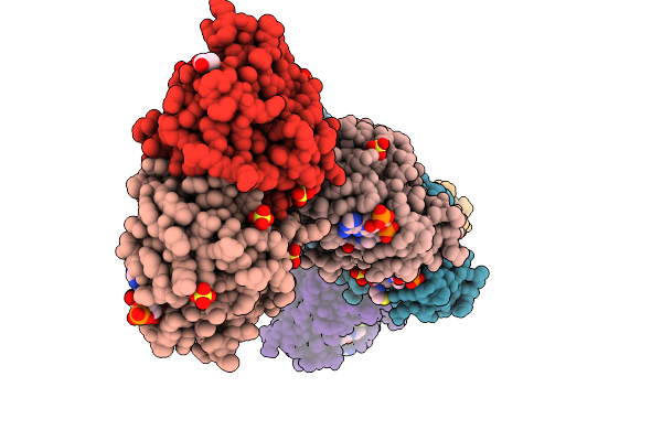 |
Crystal Structure Of N-Acetyl Transferase Domain-Containing Protein From Bacteroides Fragilis
Organism: Bacteroides fragilis
Method: X-RAY DIFFRACTION Release Date: 2025-09-10 Classification: TRANSFERASE Ligands: COA, SO4, EDO, ACT |
 |
Organism: Lama glama
Method: X-RAY DIFFRACTION Release Date: 2025-06-11 Classification: IMMUNE SYSTEM Ligands: EDO, SO4, GOL |
 |
Organism: Lama glama, Synthetic construct
Method: X-RAY DIFFRACTION Release Date: 2025-06-11 Classification: IMMUNE SYSTEM Ligands: K, EDO |
 |
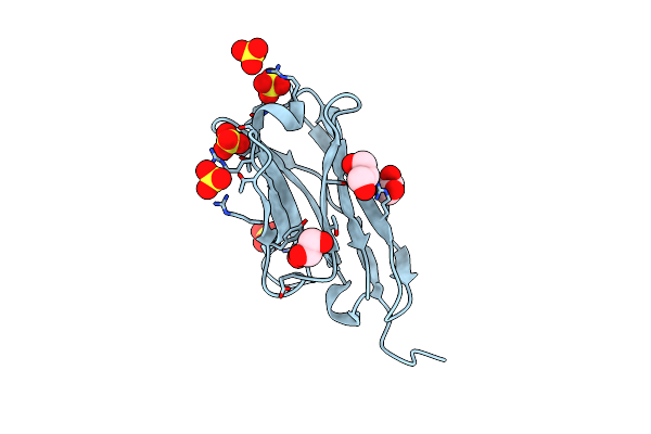
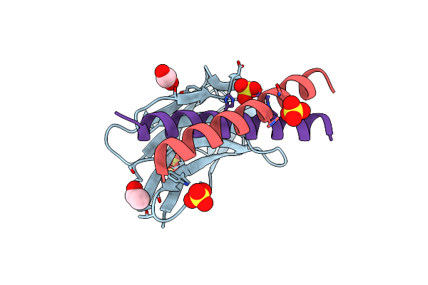
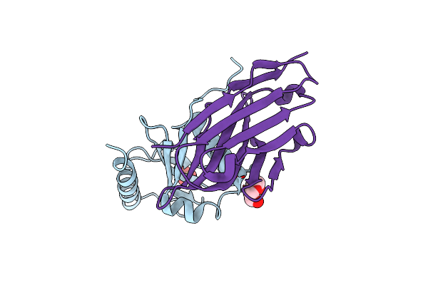
N-Terminal Domain Of Human Galectin-8 In Complex With An Alpha-Galactoside Ligand
Organism: Homo sapiens
Method: X-RAY DIFFRACTION Resolution:1.08 Å Release Date: 2025-03-12 Classification: SUGAR BINDING PROTEIN Ligands: A1IHE, CL |
 |
Organism: Vibrio cholerae
Method: X-RAY DIFFRACTION Resolution:2.33 Å Release Date: 2023-05-03 Classification: DNA BINDING PROTEIN Ligands: PO4 |
 |
Organism: Severe acute respiratory syndrome coronavirus 2
Method: X-RAY DIFFRACTION Resolution:1.93 Å Release Date: 2022-03-16 Classification: VIRAL PROTEIN Ligands: EDO, ZN |
 |
Organism: Lama glama, Synthetic construct
Method: X-RAY DIFFRACTION Resolution:1.55 Å Release Date: 2021-05-05 Classification: DE NOVO PROTEIN |
 |
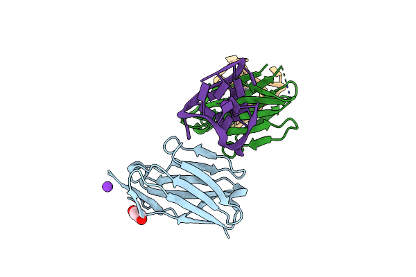
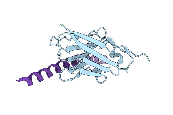
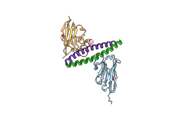
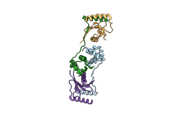
Crystal Structure Of The Aph Coiled-Coil In Complex With Nanobodies Nb28 And Nb30
Organism: Lama glama, Synthetic construct
Method: X-RAY DIFFRACTION Resolution:2.69 Å Release Date: 2021-05-05 Classification: DE NOVO PROTEIN Ligands: EDO, GOL, ACT |
 |
Organism: Lama glama, Saccharomyces cerevisiae
Method: X-RAY DIFFRACTION Resolution:2.12 Å Release Date: 2021-05-05 Classification: DE NOVO PROTEIN Ligands: SO4, ACE, EDO |
 |
Organism: Lama glama, Synthetic construct
Method: X-RAY DIFFRACTION Resolution:2.16 Å Release Date: 2021-05-05 Classification: DE NOVO PROTEIN Ligands: SO4, EDO |
 |
Organism: Lama glama, Synthetic construct
Method: X-RAY DIFFRACTION Resolution:2.00 Å Release Date: 2021-05-05 Classification: DE NOVO PROTEIN Ligands: EDO |
 |
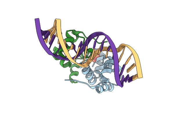
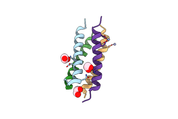
Crystal Structure Of Cbs Domain Protein From Streptococcus Pneumoniae Tigr4
Organism: Streptococcus pneumoniae serotype 4 (strain atcc baa-334 / tigr4)
Method: X-RAY DIFFRACTION Resolution:2.81 Å Release Date: 2021-03-17 Classification: STRUCTURAL GENOMICS, UNKNOWN FUNCTION Ligands: PO4 |
 |
Organism: Vibrio cholerae
Method: X-RAY DIFFRACTION Resolution:1.80 Å Release Date: 2017-04-05 Classification: ANTITOXIN |
 |
Organism: Vibrio cholerae serotype o1 (strain atcc 39315 / el tor inaba n16961), Lama glama
Method: X-RAY DIFFRACTION Resolution:2.49 Å Release Date: 2017-04-05 Classification: TOXIN Ligands: PO4, ACT, EDO, EPE, PDO |
 |
Organism: Vibrio cholerae serotype o1 (strain atcc 39315 / el tor inaba n16961), Lama glama
Method: X-RAY DIFFRACTION Resolution:1.85 Å Release Date: 2017-04-05 Classification: TOXIN Ligands: SO4, EDO |
 |
Organism: Vibrio cholerae serotype o1 (strain atcc 39315 / el tor inaba n16961)
Method: X-RAY DIFFRACTION Resolution:2.99 Å Release Date: 2017-04-05 Classification: TOXIN |
 |
Organism: Vibrio cholerae, Lama glama
Method: X-RAY DIFFRACTION Release Date: 2017-04-05 Classification: TOXIN Ligands: PEG, SO4 |
 |
Organism: Escherichia coli o157:h7 str. ss52
Method: X-RAY DIFFRACTION Resolution:3.82 Å Release Date: 2016-08-24 Classification: TOXIN |
 |
Organism: Escherichia coli o157
Method: X-RAY DIFFRACTION Resolution:2.83 Å Release Date: 2016-06-01 Classification: TOXIN Ligands: GOL |
 |
Organism: Escherichia coli o157
Method: X-RAY DIFFRACTION Resolution:2.67 Å Release Date: 2016-06-01 Classification: TOXIN Ligands: SO4 |