
Search Count: 16
 |
Organism: Pseudomonas aeruginosa
Method: X-RAY DIFFRACTION Resolution:2.65 Å Release Date: 2024-06-26 Classification: MEMBRANE PROTEIN Ligands: C8E |
 |
Organism: Pseudomonas aeruginosa
Method: X-RAY DIFFRACTION Resolution:3.96 Å Release Date: 2024-06-26 Classification: MEMBRANE PROTEIN |
 |
Organism: Pseudomonas aeruginosa
Method: X-RAY DIFFRACTION Resolution:3.10 Å Release Date: 2024-06-26 Classification: MEMBRANE PROTEIN Ligands: C8E, MG |
 |
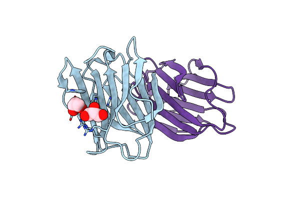
C2221 Crystal Structure Of Tama (Barrel Only) From Pseudomonas Aeruginosa At 3.15 Ang
Organism: Pseudomonas aeruginosa pao1
Method: X-RAY DIFFRACTION Resolution:3.15 Å Release Date: 2024-06-26 Classification: MEMBRANE PROTEIN Ligands: PT |
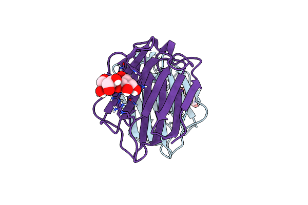 |
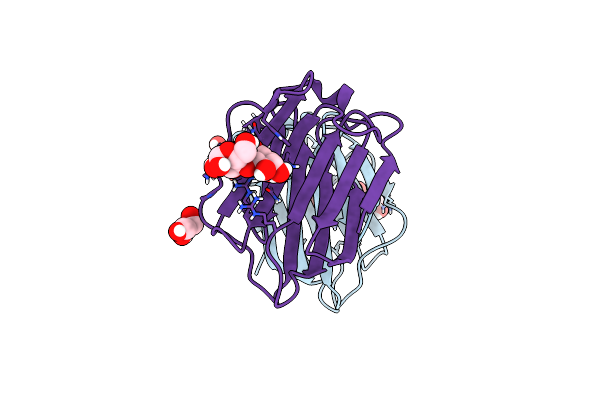
Crystal Structure Of Human Galectin-7 In Complex With 4-O-Beta-D-Galactopyranosyl-D-Glucose
Organism: Homo sapiens
Method: X-RAY DIFFRACTION Resolution:1.69 Å Release Date: 2021-08-25 Classification: SUGAR BINDING PROTEIN Ligands: LBL |
 |
Organism: Homo sapiens
Method: X-RAY DIFFRACTION Resolution:2.30 Å Release Date: 2021-08-25 Classification: SUGAR BINDING PROTEIN Ligands: GOL |
 |
Organism: Homo sapiens
Method: X-RAY DIFFRACTION Resolution:1.95 Å Release Date: 2021-08-25 Classification: SUGAR BINDING PROTEIN Ligands: GOL |
 |
Organism: Homo sapiens
Method: X-RAY DIFFRACTION Resolution:2.30 Å Release Date: 2021-08-25 Classification: SUGAR BINDING PROTEIN Ligands: TRS, GOL |
 |
Organism: Homo sapiens
Method: X-RAY DIFFRACTION Resolution:1.90 Å Release Date: 2021-08-25 Classification: SUGAR BINDING PROTEIN Ligands: GOL |
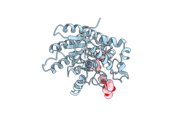 |
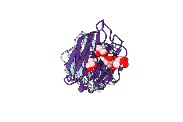
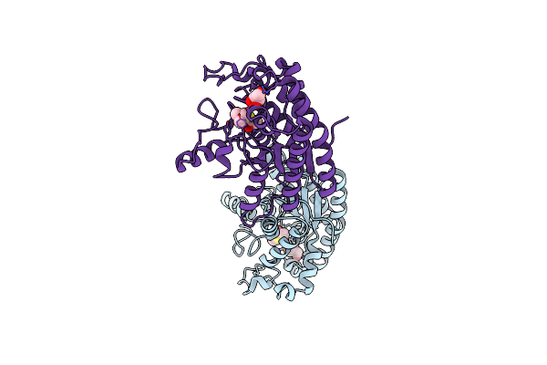
Crystal Structure Of Choe, A Bacterial Acetylcholinesterase From Pseudomonas Aeruginosa
Organism: Pseudomonas aeruginosa (strain atcc 15692 / dsm 22644 / cip 104116 / jcm 14847 / lmg 12228 / 1c / prs 101 / pao1)
Method: X-RAY DIFFRACTION Resolution:1.35 Å Release Date: 2020-05-13 Classification: HYDROLASE Ligands: MES, GOL, BUA, CL, P6G |
 |
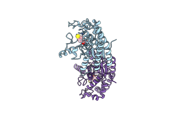
Organism: Pseudomonas aeruginosa (strain atcc 15692 / dsm 22644 / cip 104116 / jcm 14847 / lmg 12228 / 1c / prs 101 / pao1)
Method: X-RAY DIFFRACTION Resolution:1.65 Å Release Date: 2020-05-13 Classification: HYDROLASE Ligands: ACT, ETM, GOL |
 |
Organism: Pseudomonas aeruginosa (strain atcc 15692 / dsm 22644 / cip 104116 / jcm 14847 / lmg 12228 / 1c / prs 101 / pao1)
Method: X-RAY DIFFRACTION Resolution:1.85 Å Release Date: 2020-05-13 Classification: HYDROLASE Ligands: QFJ, IOD, PPI, ETM |
 |
Organism: Pseudomonas aeruginosa (strain atcc 15692 / dsm 22644 / cip 104116 / jcm 14847 / lmg 12228 / 1c / prs 101 / pao1)
Method: X-RAY DIFFRACTION Resolution:1.57 Å Release Date: 2020-05-13 Classification: HYDROLASE Ligands: AT3, GOL |
 |
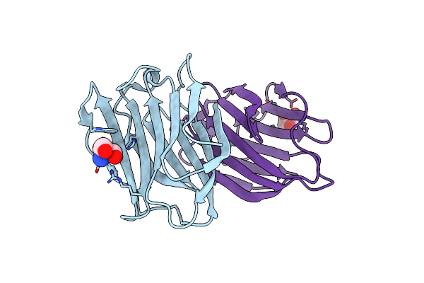
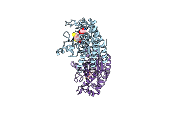
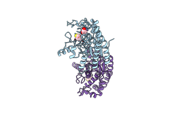
Crystal Structure Of Choe D285N Mutant In Complex With Acetate And Thiocholine
Organism: Pseudomonas aeruginosa (strain atcc 15692 / dsm 22644 / cip 104116 / jcm 14847 / lmg 12228 / 1c / prs 101 / pao1)
Method: X-RAY DIFFRACTION Resolution:1.43 Å Release Date: 2020-05-13 Classification: HYDROLASE Ligands: ACT, ETM |
 |
Organism: Pseudomonas aeruginosa (strain atcc 15692 / dsm 22644 / cip 104116 / jcm 14847 / lmg 12228 / 1c / prs 101 / pao1)
Method: X-RAY DIFFRACTION Resolution:1.80 Å Release Date: 2020-05-13 Classification: HYDROLASE Ligands: ETM, GOL |
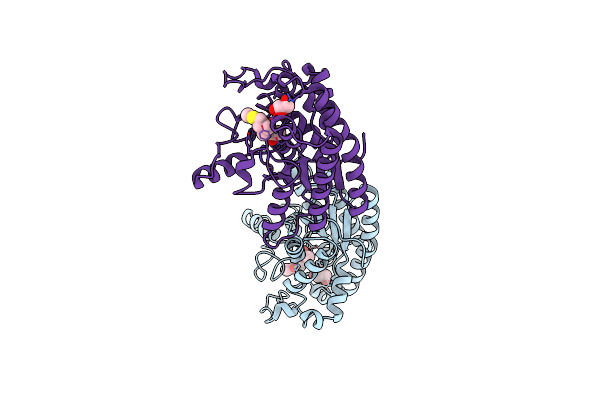 |
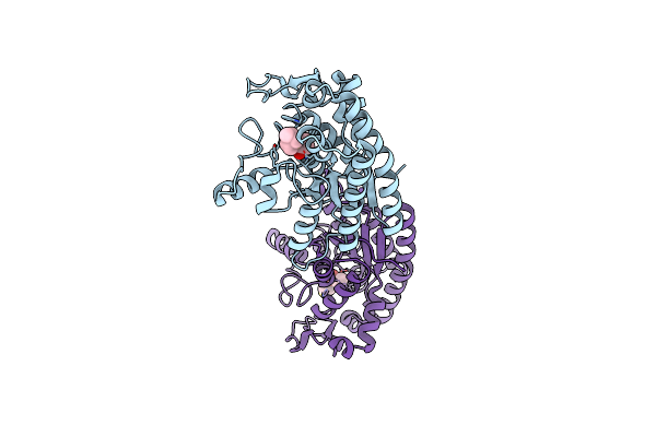
Crystal Structure Of Choe S38A Mutant In Complex With Acetate And Acetylthiocholine
Organism: Pseudomonas aeruginosa (strain atcc 15692 / dsm 22644 / cip 104116 / jcm 14847 / lmg 12228 / 1c / prs 101 / pao1)
Method: X-RAY DIFFRACTION Resolution:1.42 Å Release Date: 2020-05-13 Classification: HYDROLASE Ligands: ACT, AT3, GOL |