
Search Count: 6
 |
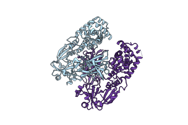
Organism: Corynebacterium diphtheriae
Method: X-RAY DIFFRACTION Resolution:2.25 Å Release Date: 2023-07-05 Classification: TOXIN |
 |
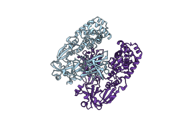
Organism: Corynebacterium diphtheriae
Method: X-RAY DIFFRACTION Resolution:2.10 Å Release Date: 2023-07-05 Classification: TOXIN Ligands: APU |
 |
Organism: Corynebacterium diphtheriae
Method: X-RAY DIFFRACTION Resolution:2.05 Å Release Date: 2020-11-18 Classification: TOXIN |
 |
Organism: Corynebacterium diphtheriae
Method: X-RAY DIFFRACTION Resolution:2.05 Å Release Date: 2020-11-18 Classification: TOXIN |
 |
Organism: Corynebacterium diphtheriae
Method: X-RAY DIFFRACTION Resolution:2.10 Å Release Date: 2020-11-18 Classification: TOXIN |
 |
Organism: Corynebacterium diphtheriae
Method: X-RAY DIFFRACTION Resolution:2.30 Å Release Date: 2020-11-18 Classification: TOXIN |