
Search Count: 22
 |
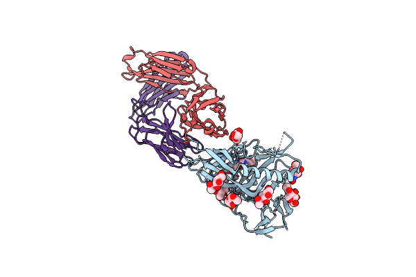
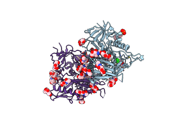
Organism: Human immunodeficiency virus 1
Method: X-RAY DIFFRACTION Resolution:2.70 Å Release Date: 2016-03-30 Classification: VIRAL PROTEIN Ligands: 5VE |
 |
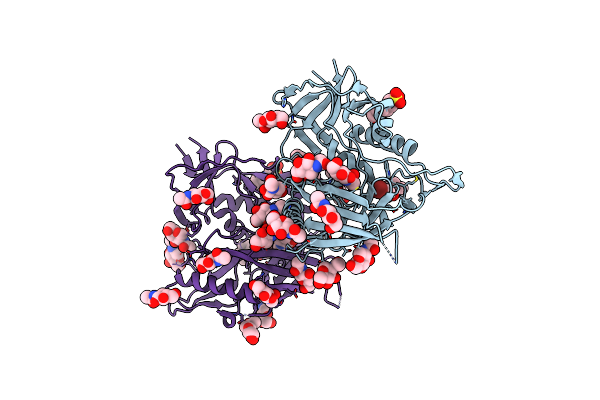
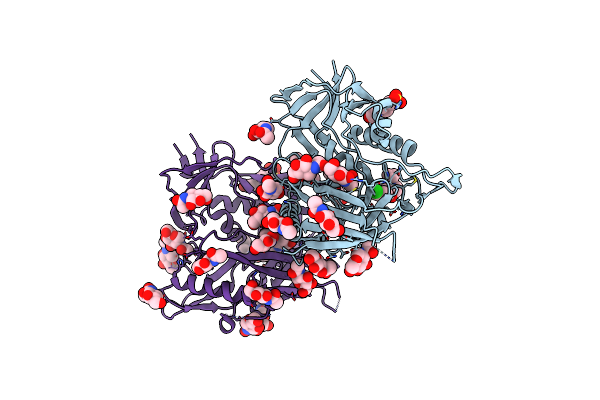
Organism: Human immunodeficiency virus 1
Method: X-RAY DIFFRACTION Resolution:2.60 Å Release Date: 2016-03-30 Classification: VIRAL PROTEIN Ligands: NAG, 5VG |
 |
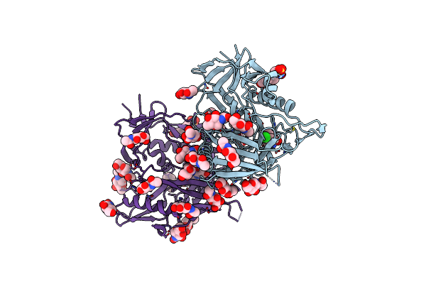
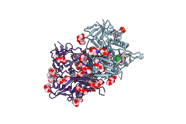
Organism: Human immunodeficiency virus 1
Method: X-RAY DIFFRACTION Resolution:2.80 Å Release Date: 2016-03-30 Classification: VIRAL PROTEIN Ligands: FMT, NAG, 5VF |
 |
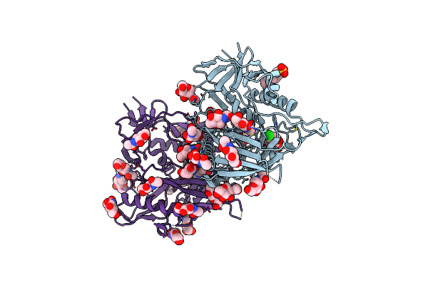
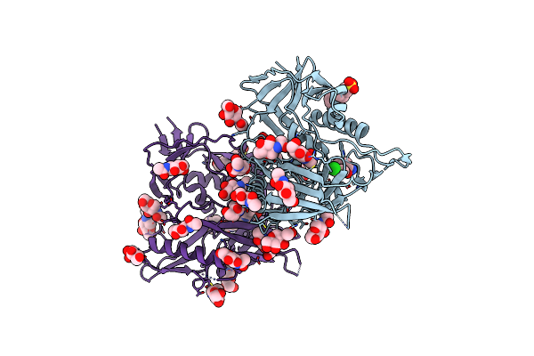
Organism: Human immunodeficiency virus 1
Method: X-RAY DIFFRACTION Resolution:3.10 Å Release Date: 2016-03-30 Classification: VIRAL PROTEIN Ligands: NAG, 5VH |
 |
Crystal Structure Of Clade C1086 Hiv-1 Gp120 Core In Complex With Dmj-Ii-121
Organism: Human immunodeficiency virus type 1
Method: X-RAY DIFFRACTION Resolution:2.50 Å Release Date: 2013-05-29 Classification: VIRAL PROTEIN/INHIBITOR Ligands: 1C1, NAG, FMT |
 |
Crystal Structure Of Clade A/E 93Th057 Hiv-1 Gp120 H375S Core In Complex With Dmj-Ii-121
Organism: Human immunodeficiency virus type 1
Method: X-RAY DIFFRACTION Resolution:2.50 Å Release Date: 2013-05-29 Classification: VIRAL PROTEIN/INHIBITOR Ligands: 1C1, EPE, NAG |
 |
Organism: Human immunodeficiency virus type 1 group m subtype b (isolate yu-2), Homo sapiens
Method: X-RAY DIFFRACTION Resolution:2.50 Å Release Date: 2013-02-27 Classification: IMMUNE SYSTEM/TRANSCRIPTION INHIBITOR Ligands: NAG, 0LY |
 |
Crystal Structure Of Clade A/E 93Th057 Hiv-1 Gp120 Core In Complex With Nbd-557
Organism: Human immunodeficiency virus 1
Method: X-RAY DIFFRACTION Resolution:2.10 Å Release Date: 2013-02-27 Classification: viral protein/transcription inhibitor Ligands: NAG, 0LY, EPE |
 |
Crystal Structure Of Clade A/E 93Th057 Hiv-1 Gp120 Core In Complex With As-Ii-37
Organism: Human immunodeficiency virus 1
Method: X-RAY DIFFRACTION Resolution:2.40 Å Release Date: 2013-02-27 Classification: viral protein/transcription inhibitor Ligands: NAG, 0LZ, EPE |
 |
Crystal Structure Of Clade A/E 93Th057 Hiv-1 Gp120 Core In Complex With As-I-261
Organism: Human immunodeficiency virus 1
Method: X-RAY DIFFRACTION Resolution:1.94 Å Release Date: 2013-02-27 Classification: viral protein/transcription inhibitor Ligands: NAG, EPE, 0M1 |
 |
Crystal Structure Of Clade A/E 93Th057 Hiv-1 Gp120 Core In Complex With Mae-Ii-167
Organism: Human immunodeficiency virus 1
Method: X-RAY DIFFRACTION Resolution:2.20 Å Release Date: 2013-02-27 Classification: viral protein/transcription inhibitor Ligands: NAG, EPE, 0M4 |
 |
Crystal Structure Of Clade A/E 93Th057 Hiv-1 Gp120 Core In Complex With Mae-Ii-188
Organism: Human immunodeficiency virus 1
Method: X-RAY DIFFRACTION Resolution:2.40 Å Release Date: 2013-02-27 Classification: viral protein/transcription inhibitor Ligands: NAG, 0M5, EPE |
 |
Crystal Structure Of Clade A/E 93Th057 Hiv-1 Gp120 Core In Complex With Ts-Ii-224
Organism: Human immunodeficiency virus type 1
Method: X-RAY DIFFRACTION Resolution:1.98 Å Release Date: 2012-05-02 Classification: VIRAL PROTEIN/INHIBITOR Ligands: NAG, EPE, 0LM |
 |
Crystal Structure Of Clade A/E 93Th057 Hiv-1 Gp120 Core In Complex With Aws-I-50
Organism: Human immunodeficiency virus type 1
Method: X-RAY DIFFRACTION Resolution:1.80 Å Release Date: 2012-05-02 Classification: VIRAL PROTEIN/INHIBITOR Ligands: NAG, EPE, 0LL |
 |
Crystal Structure Of Clade A/E 93Th057 Hiv-1 Gp120 Core In Complex With Dmj-I-228
Organism: Human immunodeficiency virus type 1
Method: X-RAY DIFFRACTION Resolution:1.89 Å Release Date: 2012-05-02 Classification: VIRAL PROTEIN/INHIBITOR Ligands: NAG, EPE, 0LK |
 |
Crystal Structure Of Clade A/E 93Th057 Hiv-1 Gp120 Core In Complex With Aws-I-169
Organism: Human immunodeficiency virus type 1
Method: X-RAY DIFFRACTION Resolution:1.80 Å Release Date: 2012-05-02 Classification: VIRAL PROTEIN/INHIBITOR Ligands: NAG, EPE, 0LJ |
 |
Organism: Homo sapiens
Method: X-RAY DIFFRACTION Resolution:2.60 Å Release Date: 1999-10-24 Classification: HYDROLASE |
 |
Use Of Papain As A Model For The Structure-Based Design Of Cathepsin K Inhibitors. Crystal Structures Of Two Papain Inhibitor Complexes Demonstrate Binding To S'-Subsites.
Organism: Carica papaya
Method: X-RAY DIFFRACTION Resolution:2.50 Å Release Date: 1999-08-16 Classification: HYDROLASE Ligands: SBA |
 |
Use Of Papain As A Model For The Structure-Based Design Of Cathepsin K Inhibitors. Crystal Structures Of Two Papain Inhibitor Complexes Demonstrate Binding To S'-Subsites.
Organism: Carica papaya
Method: X-RAY DIFFRACTION Resolution:2.20 Å Release Date: 1999-08-12 Classification: HYDROLASE Ligands: ALD |
 |
Adipocyte Lipid Binding Protein Complexed With Arachidonic Acid: X-Ray Crystallographic And Titration Calorimetry Studies
Organism: Mus musculus
Method: X-RAY DIFFRACTION Resolution:1.60 Å Release Date: 1994-12-20 Classification: LIPID BINDING PROTEIN Ligands: ACD, PPI |