
Search Count: 42
 |
Organism: Measles morbillivirus, Mus musculus
Method: ELECTRON MICROSCOPY Release Date: 2024-07-03 Classification: VIRAL PROTEIN Ligands: NAG |
 |
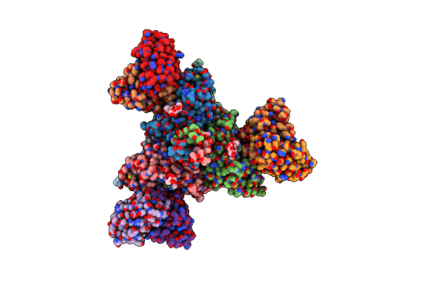
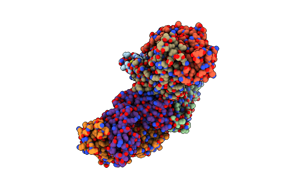
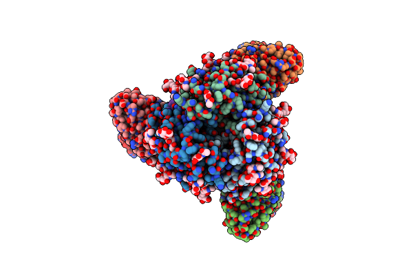
Structure Of The Measles Virus Fusion Protein In The Post-Fusion Conformation
Organism: Measles virus strain ichinose-b95a
Method: ELECTRON MICROSCOPY Release Date: 2024-07-03 Classification: VIRAL PROTEIN Ligands: NAG |
 |
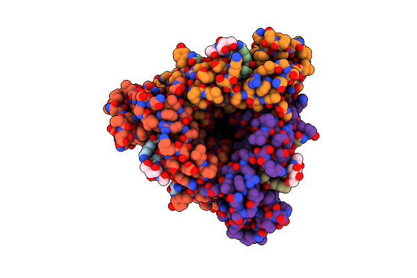
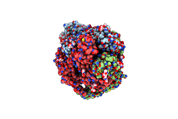
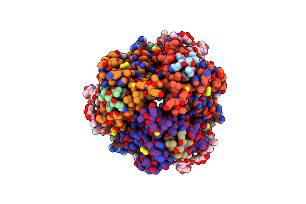
Structure Of The Measles Virus Fusion Protein In The Pre-Fusion Conformation
Organism: Measles virus strain ichinose-b95a
Method: ELECTRON MICROSCOPY Release Date: 2024-07-03 Classification: VIRAL PROTEIN Ligands: NAG |
 |
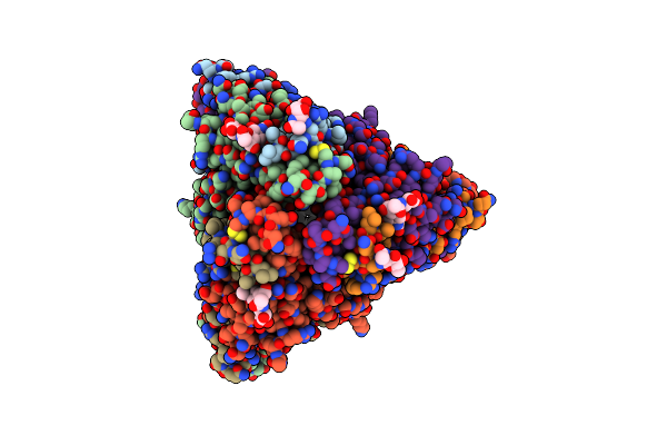
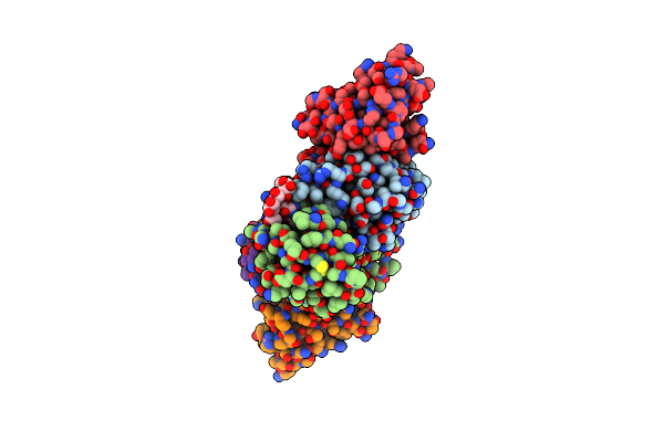
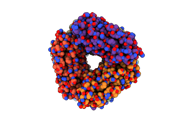
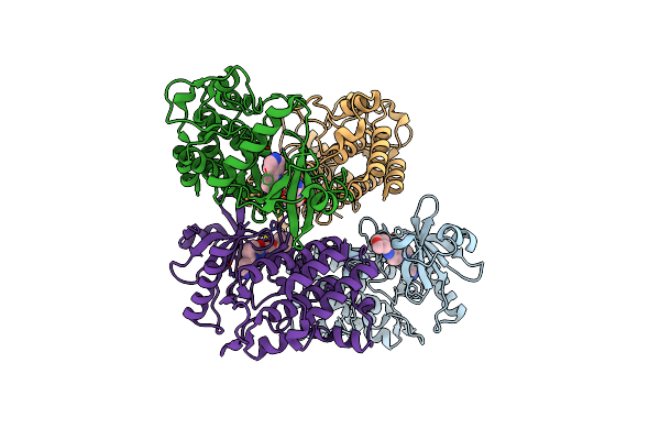
Structure Of The Measles Virus Fusion Protein In The Pre-Fusion Conformation With Bound [Fip-Hrc]2-Peg11
Organism: Measles virus strain ichinose-b95a, Synthetic construct
Method: ELECTRON MICROSCOPY Release Date: 2024-07-03 Classification: VIRAL PROTEIN Ligands: NAG |
 |
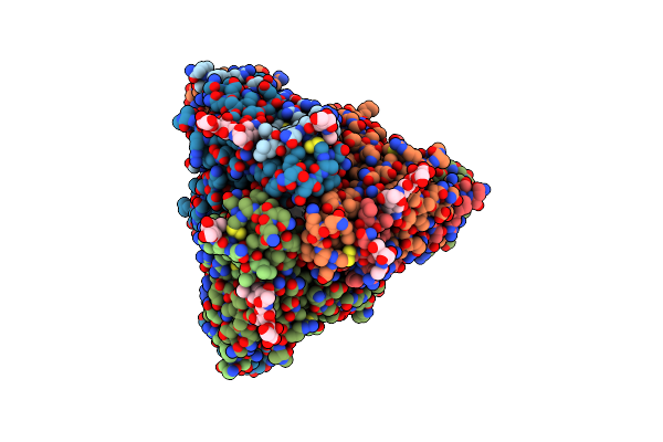
Organism: Measles virus strain ichinose-b95a, Homo sapiens
Method: ELECTRON MICROSCOPY Release Date: 2024-07-03 Classification: VIRAL PROTEIN Ligands: NAG |
 |
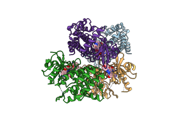
Organism: Escherichia coli
Method: ELECTRON MICROSCOPY Release Date: 2024-04-10 Classification: STRUCTURAL PROTEIN |
 |
2.5 A Cryo-Em Structure Of The In Vitro Fimd-Catalyzed Assembly Of Type 1 Pilus Rod
Organism: Escherichia coli
Method: ELECTRON MICROSCOPY Release Date: 2024-04-10 Classification: STRUCTURAL PROTEIN |
 |
Organism: Homo sapiens, Severe acute respiratory syndrome coronavirus 2
Method: X-RAY DIFFRACTION Resolution:2.57 Å Release Date: 2023-06-28 Classification: VIRAL PROTEIN Ligands: NAG |
 |
Structure Of Sars-Cov-2 Omicron Ba.1 Spike In Complex With Antibody Fab 1C3
Organism: Homo sapiens, Severe acute respiratory syndrome coronavirus 2
Method: ELECTRON MICROSCOPY Release Date: 2023-05-03 Classification: VIRAL PROTEIN/IMMUNE SYSTEM Ligands: NAG |
 |
Structure Of Sars-Cov-2 Spike With Antibody Fabs 2A10 And 1H2 (Local Refinement Of The Rbd And Fabs 1H2 And 2A10)
Organism: Homo sapiens, Severe acute respiratory syndrome coronavirus 2
Method: ELECTRON MICROSCOPY Release Date: 2023-05-03 Classification: VIRAL PROTEIN/IMMUNE SYSTEM |
 |
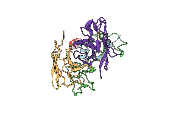
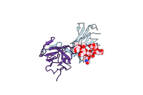
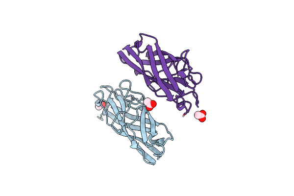
Lymphocytic Choriomeningitis Virus Glycoprotein In Complex With Neutralizing Antibody M28
Organism: Lymphocytic choriomeningitis virus, Homo sapiens
Method: ELECTRON MICROSCOPY Release Date: 2023-04-12 Classification: VIRAL PROTEIN/IMMUNE SYSTEM Ligands: NAG |
 |
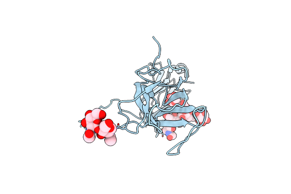
Organism: Lymphocytic choriomeningitis virus
Method: ELECTRON MICROSCOPY Release Date: 2023-04-12 Classification: VIRAL PROTEIN Ligands: NAG |
 |
Organism: Escherichia coli
Method: ELECTRON MICROSCOPY Resolution:2.85 Å Release Date: 2021-03-31 Classification: STRUCTURAL PROTEIN |
 |
Organism: Homo sapiens
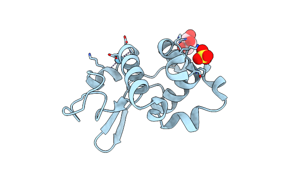
Method: X-RAY DIFFRACTION Resolution:2.44 Å Release Date: 2020-11-25 Classification: TRANSFERASE/TRANSFERASE INHIBITOR Ligands: FJ0 |
 |
Crystal Structure Of Irak4 Kinase In Complex With A Small Molecule Inhibitor
Organism: Homo sapiens
Method: X-RAY DIFFRACTION Resolution:3.20 Å Release Date: 2020-11-25 Classification: TRANSFERASE/TRANSFERASE INHIBITOR Ligands: FJ9 |
 |
Organism: Homo sapiens
Method: ELECTRON MICROSCOPY Release Date: 2020-09-02 Classification: ANTIMICROBIAL PROTEIN |
 |
Organism: Homo sapiens
Method: ELECTRON MICROSCOPY Release Date: 2020-09-02 Classification: ANTIMICROBIAL PROTEIN |
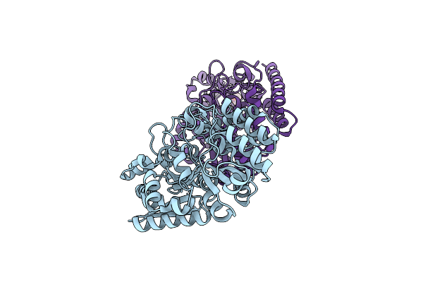 |
Sn-Glycerol-3-Phosphate Binding Periplasmic Protein Ugpb From Escherichia Coli - W169S, W172S
Organism: Escherichia coli (strain k12)
Method: X-RAY DIFFRACTION Resolution:1.25 Å Release Date: 2020-08-19 Classification: CHAPERONE Ligands: GOL |
 |
Organism: Bos taurus
Method: X-RAY DIFFRACTION Resolution:1.85 Å Release Date: 2019-02-20 Classification: METAL BINDING PROTEIN Ligands: SO4, GOL, LA |
 |
Organism: Salmonella paratyphi a (strain atcc 9150 / sarb42)
Method: X-RAY DIFFRACTION Resolution:1.69 Å Release Date: 2019-01-30 Classification: STRUCTURAL PROTEIN Ligands: ACY |