
Search Count: 17
 |
Organism: Synthetic construct
Method: SOLUTION NMR Release Date: 2021-12-01 Classification: DNA Ligands: LU |
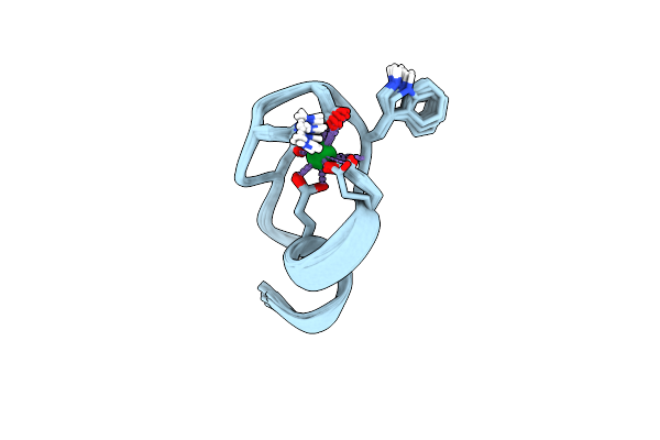 |
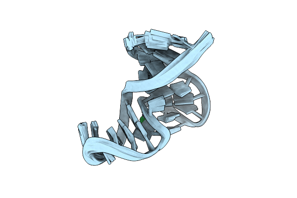
The Binding Structure Of A Lanthanide Binding Tag (Lbt3) With Lutetium Ion (Lu3+)
Organism: Homo sapiens
Method: SOLUTION NMR Release Date: 2021-04-28 Classification: DE NOVO PROTEIN Ligands: LU |
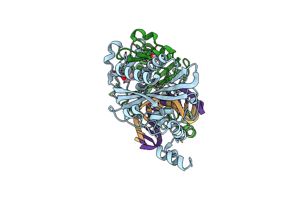 |
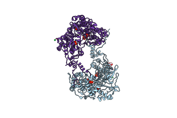
Crystal Structure Of The Ternary Ecorv-Dna-Lu Complex With Uncleaved Dna Substrate. Lanthanide Binding To Ecorv-Dna Complex Inhibits Cleavage.
Organism: Escherichia coli, Synthetic construct
Method: X-RAY DIFFRACTION Resolution:1.76 Å Release Date: 2016-11-09 Classification: HYDROLASE/DNA Ligands: LU, NA, EDO |
 |
Crystal Structure Of The Ternary Ecorv-Dna-Lu Complex With Cleaved Dna Substrate.
Organism: Escherichia coli, Synthetic construct
Method: X-RAY DIFFRACTION Resolution:2.00 Å Release Date: 2016-11-09 Classification: HYDROLASE/DNA Ligands: EDO, NA, LU |
 |
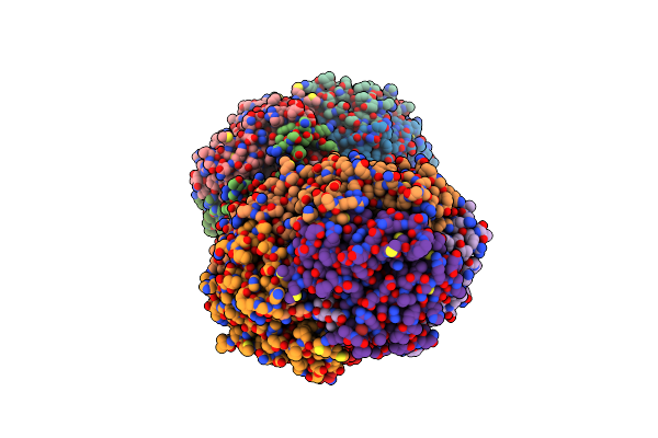
Crystal Structure Of Thosea Asigna Virus Rna-Dependent Rna Polymerase (Rdrp) Complexed With Lu3+
Organism: Thosea asigna virus
Method: X-RAY DIFFRACTION Resolution:3.00 Å Release Date: 2015-11-04 Classification: TRANSFERASE Ligands: LU, SO4, GOL |
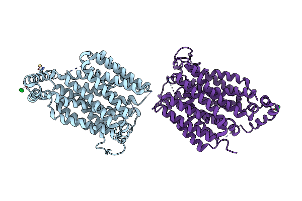 |
Partially Occluded Inward Open Conformation Of The Xylose Transporter Xyle From E. Coli
Organism: Escherichia coli
Method: X-RAY DIFFRACTION Resolution:3.80 Å Release Date: 2013-05-01 Classification: TRANSPORT PROTEIN Ligands: CD, LU |
 |
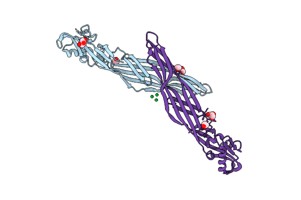
Organism: Mus musculus, Eptatretus burgeri
Method: X-RAY DIFFRACTION Resolution:2.40 Å Release Date: 2009-11-24 Classification: IMMUNE SYSTEM Ligands: NAG, PDK, LU |
 |
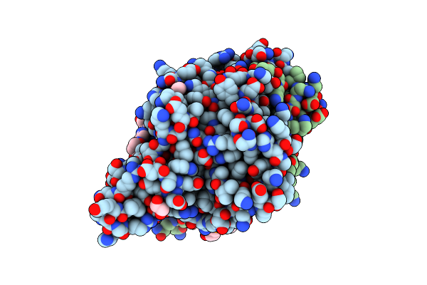
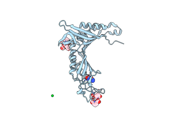
Crystal Structure Of The Parasporin-2 Bacillus Thuringiensis Toxin That Recognizes Cancer Cells
Organism: Bacillus thuringiensis serovar dakota
Method: X-RAY DIFFRACTION Resolution:2.38 Å Release Date: 2009-01-13 Classification: TOXIN Ligands: LU, GOL, EDO, CL |
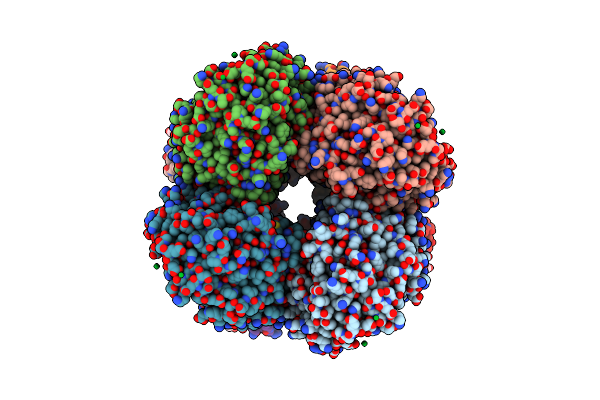 |
Structure Of The Octameric Penicillin-Binding Protein Homologue From Pyrococcus Abyssi
Organism: Pyrococcus abyssi
Method: X-RAY DIFFRACTION Resolution:2.20 Å Release Date: 2008-07-22 Classification: HYDROLASE Ligands: LU, DO3 |
 |
Organism: Aspergillus flavus
Method: X-RAY DIFFRACTION Resolution:1.60 Å Release Date: 2008-04-22 Classification: OXIDOREDUCTASE Ligands: LU, AZA, PDC |
 |
Organism: Clarkia breweri
Method: X-RAY DIFFRACTION Resolution:3.00 Å Release Date: 2003-09-09 Classification: TRANSFERASE Ligands: LU, SAH, SAL |
 |
Organism: Homo sapiens
Method: X-RAY DIFFRACTION Resolution:2.90 Å Release Date: 2003-06-17 Classification: OXIDOREDUCTASE Ligands: LU, NAD |
 |
Organism: Bos taurus
Method: X-RAY DIFFRACTION Resolution:2.00 Å Release Date: 2003-01-07 Classification: STRUCTURAL PROTEIN Ligands: BR, GOL, LU |
 |
Crystal Structure Of Rabbit Hemorrhagic Disease Virus Rna-Dependent Rna Polymerase Complexed With Lu3+
Organism: Rabbit hemorrhagic disease virus
Method: X-RAY DIFFRACTION Resolution:2.50 Å Release Date: 2002-01-16 Classification: TRANSFERASE Ligands: LU |
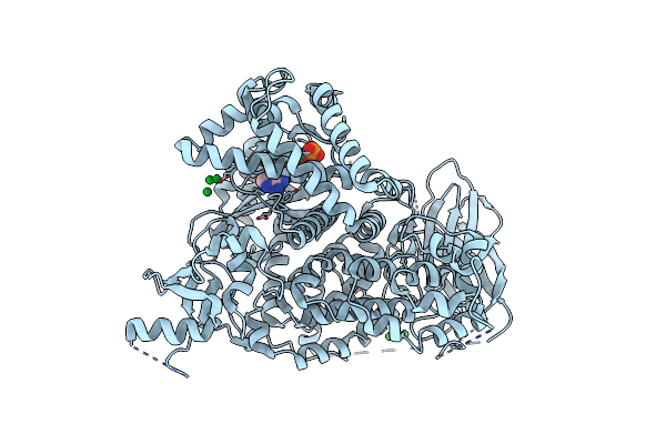 |
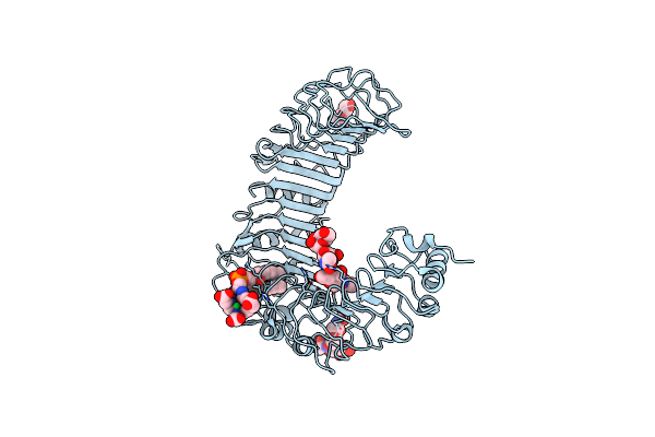
Structural Insights Into Phoshoinositide 3-Kinase Enzymatic Mechanism And Signalling
Organism: Sus scrofa
Method: X-RAY DIFFRACTION Resolution:2.20 Å Release Date: 2000-10-26 Classification: PHOSPHOINOSITIDE 3-KINASE GAMMA Ligands: ATP, LU |
 |
Method: X-RAY DIFFRACTION
Resolution:2.70 Å Release Date: 2000-05-08 Classification: RNA Ligands: LU, MG, SO4 |
 |
Pectate Lyase C From Erwinia Chrysanthemi With 1 Lu+3 Ion In The Putative Calcium Binding Site
Organism: Erwinia chrysanthemi
Method: X-RAY DIFFRACTION Resolution:2.20 Å Release Date: 1999-07-13 Classification: LYASE Ligands: LU |