
Search Count: 12
All
Selected
 |
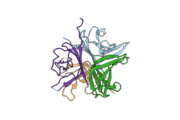
Topological Model Of The P2 Virion Baseplate In Activated Conformation (Closed Tal Trimer)
Organism: Lactococcus phage p2
Method: ELECTRON MICROSCOPY Release Date: 2020-08-26 Classification: VIRAL PROTEIN |
 |
Organism: Lactococcus phage p2
Method: ELECTRON MICROSCOPY Release Date: 2020-08-26 Classification: VIRAL PROTEIN |
 |
Organism: Lactococcus phage p2
Method: ELECTRON MICROSCOPY Release Date: 2020-08-26 Classification: VIRAL PROTEIN |
 |
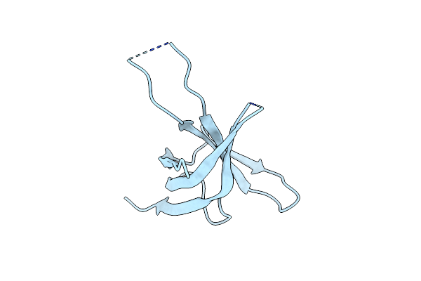
Organism: Lactococcus phage p2
Method: SOLUTION NMR Release Date: 2020-04-01 Classification: VIRAL PROTEIN |
 |
Organism: Lactococcus phage p2
Method: X-RAY DIFFRACTION Resolution:5.46 Å Release Date: 2014-07-09 Classification: VIRAL PROTEIN Ligands: CA |
 |
Structures Of Lactococcal Phage P2 Baseplate Shed Light On A Novel Mechanism Of Host Attachment And Activation In Siphoviridae
Organism: Lactococcus phage p2, Lama glama
Method: X-RAY DIFFRACTION Resolution:2.60 Å Release Date: 2010-02-16 Classification: VIRAL PROTEIN |
 |
Organism: Lactococcus phage p2
Method: X-RAY DIFFRACTION Resolution:3.90 Å Release Date: 2010-02-16 Classification: VIRAL PROTEIN Ligands: SR |
 |
Crystal Structure From A Single-Stranded Dna Binding Protein From The Lactococcal Phage P2
Organism: Lactococcus phage p2
Method: X-RAY DIFFRACTION Resolution:2.60 Å Release Date: 2009-09-15 Classification: DNA BINDING PROTEIN |
 |
Crystal Structure Of A Double Ile-To-Met Mutant Of Protein Orf34 From Lactococcus Phage P2
Organism: Lactococcus phage p2
Method: X-RAY DIFFRACTION Resolution:2.10 Å Release Date: 2009-09-15 Classification: DNA BINDING PROTEIN |
 |
Crystal Structure Of A Cleaved Form Of A Chimeric Receptor Binding Protein From Lactococcal Phages Subspecies Tp901-1 And P2
Organism: Lactococcus phage tp901-1, Lactococcus phage p2
Method: X-RAY DIFFRACTION Resolution:1.65 Å Release Date: 2009-06-09 Classification: VIRAL PROTEIN |
 |
Organism: Lactococcus phage p2
Method: X-RAY DIFFRACTION Resolution:2.90 Å Release Date: 2009-04-14 Classification: VIRAL PROTEIN |
 |
Crystal Structure Of A Chimeric Receptor Binding Protein From Lactococcal Phages Subspecies Tp901-1 And P2
Organism: Lactococcus phage tp901-1, Lactococcus phage p2
Method: X-RAY DIFFRACTION Resolution:3.35 Å Release Date: 2009-04-14 Classification: VIRUS/VIRAL PROTEIN |