
Search Count: 232
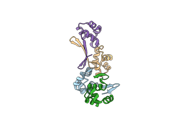 |
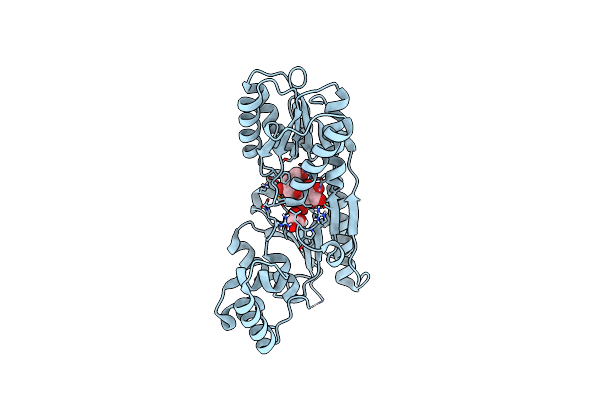
Organism: Mycobacterium tuberculosis (strain atcc 25618 / h37rv)
Method: X-RAY DIFFRACTION Resolution:2.05 Å Release Date: 2022-03-02 Classification: DNA BINDING PROTEIN |
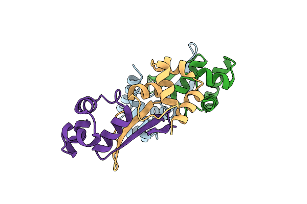 |
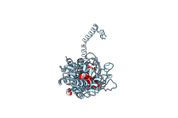
Organism: Mycobacterium tuberculosis (strain atcc 25618 / h37rv)
Method: X-RAY DIFFRACTION Resolution:3.20 Å Release Date: 2022-03-02 Classification: DNA BINDING PROTEIN |
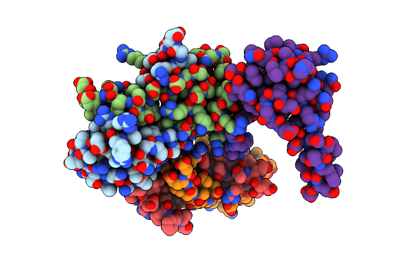 |
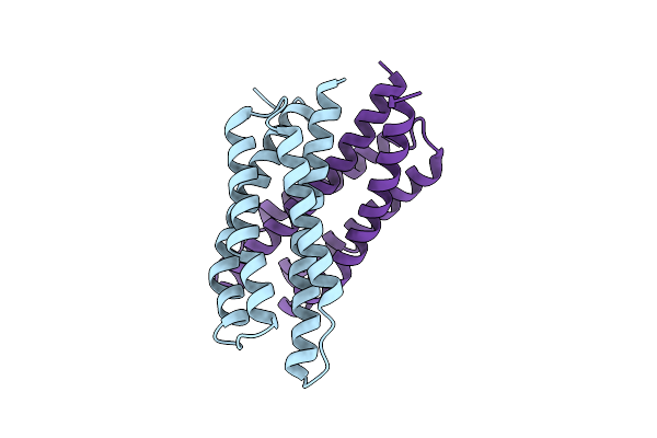
Organism: Mycobacterium tuberculosis (strain atcc 25618 / h37rv)
Method: X-RAY DIFFRACTION Resolution:3.41 Å Release Date: 2022-03-02 Classification: DNA BINDING PROTEIN |
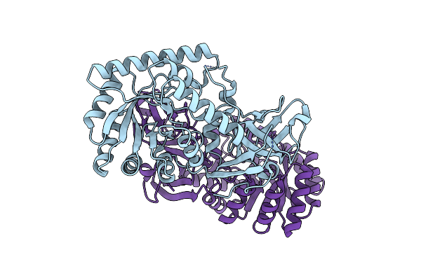 |
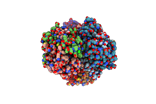
Organism: Candidatus filomicrobium marinum
Method: X-RAY DIFFRACTION Resolution:2.20 Å Release Date: 2021-08-04 Classification: BIOSYNTHETIC PROTEIN |
 |
Organism: Mus musculus
Method: ELECTRON MICROSCOPY Release Date: 2021-03-10 Classification: GENE REGULATION |
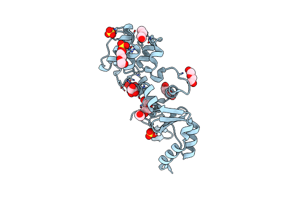 |
The Structure Of A Sensor Domain Of A Histidine Kinase (Vxra) From Vibrio Cholerae O1 Biovar Eltor Str. N16961, N239 Deletion Mutant
Organism: Vibrio cholerae serotype o1 (strain atcc 39315 / el tor inaba n16961)
Method: X-RAY DIFFRACTION Resolution:1.98 Å Release Date: 2021-01-27 Classification: SIGNALING PROTEIN Ligands: GOL, PEG, PG4, SO4 |
 |
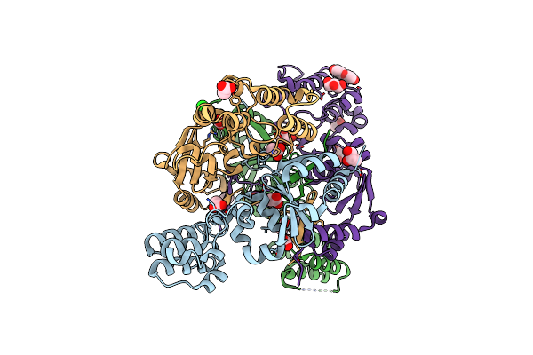
Crystal Structure Of Covid-19 Virus Spike Receptor-Binding Domain Complexed With A Neutralizing Antibody Ct-P59
Organism: Severe acute respiratory syndrome coronavirus 2, Homo sapiens
Method: X-RAY DIFFRACTION Resolution:2.71 Å Release Date: 2021-01-20 Classification: VIRAL PROTEIN Ligands: NI, EDO |
 |
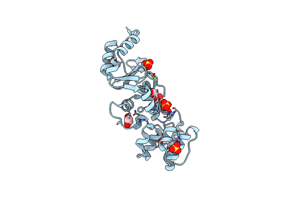
The Structure Of A Sensor Domain Of A Histidine Kinase (Vxra) From Vibrio Cholerae O1 Biovar Eltor Str. N16961, 2Nd Form
Organism: Vibrio cholerae serotype o1 (strain atcc 39315 / el tor inaba n16961)
Method: X-RAY DIFFRACTION Resolution:2.25 Å Release Date: 2020-10-14 Classification: SIGNALING PROTEIN Ligands: GOL, ACT, CL, PEG, SRT |
 |
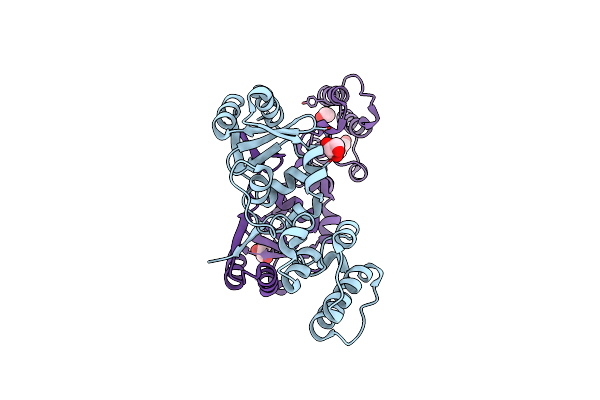
The Structure Of A Sensor Domain Of A Histidine Kinase (Vxra) From Vibrio Cholerae O1 Biovar Eltor Str. N16961, N239-T240 Deletion Mutant
Organism: Vibrio cholerae serotype o1 (strain atcc 39315 / el tor inaba n16961)
Method: X-RAY DIFFRACTION Resolution:2.20 Å Release Date: 2020-10-14 Classification: SIGNALING PROTEIN Ligands: EDO, SO4, MG |
 |
The Structure Of A Sensor Domain Of A Histidine Kinase (Vxra) From Vibrio Cholerae O1 Biovar Eltor Str. N16961, D238-T240 Deletion Mutant
Organism: Vibrio cholerae serotype o1 (strain atcc 39315 / el tor inaba n16961)
Method: X-RAY DIFFRACTION Resolution:1.98 Å Release Date: 2020-10-14 Classification: SIGNALING PROTEIN Ligands: GOL, EDO |
 |
2.1 Angstrom Resolution Crystal Structure Of 5'-Methylthioadenosine/S-Adenosylhomocysteine Nucleosidase (Mtnn) From Haemophilus Influenzae Pittii.
Organism: Haemophilus influenzae pittii
Method: X-RAY DIFFRACTION Resolution:2.10 Å Release Date: 2019-07-17 Classification: HYDROLASE Ligands: HCS, ADE, BO3 |
 |
1.3 Angstrom Resolution Crystal Structure Of Udp-N-Acetylglucosamine 1-Carboxyvinyltransferase From Streptococcus Pneumoniae In Complex With (2R)-2-(Phosphonooxy)Propanoic Acid.
Organism: Streptococcus pneumoniae serotype 4 (strain atcc baa-334 / tigr4)
Method: X-RAY DIFFRACTION Resolution:1.30 Å Release Date: 2019-01-16 Classification: TRANSFERASE Ligands: 0V5, CIT, CL, EDO |
 |
1.7 Angstrom Resolution Crystal Structure Of Arginase From Bacillus Subtilis Subsp. Subtilis Str. 168
Organism: Bacillus subtilis (strain 168)
Method: X-RAY DIFFRACTION Resolution:1.70 Å Release Date: 2019-01-02 Classification: HYDROLASE Ligands: CL, NA, MG, FMT, URE, EDO, SO4, PEG, GOA |
 |
Organism: Citrobacter freundii
Method: X-RAY DIFFRACTION Resolution:2.28 Å Release Date: 2018-10-31 Classification: OXIDOREDUCTASE Ligands: NAD |
 |
Organism: Thermobifida fusca yx
Method: X-RAY DIFFRACTION Resolution:1.63 Å Release Date: 2018-08-22 Classification: TRANSCRIPTION Ligands: GOL |
 |
1.50 Angstrom Resolution Crystal Structure Of Argininosuccinate Synthase From Bordetella Pertussis In Complex With Amp.
Organism: Bordetella pertussis
Method: X-RAY DIFFRACTION Resolution:1.50 Å Release Date: 2018-08-01 Classification: HYDROLASE Ligands: AMP, CL, SO4, EDO, MLA |
 |
2.0 Angstrom Resolution Crystal Structure Of N-Terminal Ligand-Binding Domain Of Putative Methyl-Accepting Chemotaxis Protein From Salmonella Enterica
Organism: Salmonella enterica subsp. enterica serovar typhimurium str. lt2
Method: X-RAY DIFFRACTION Resolution:2.00 Å Release Date: 2018-05-16 Classification: TRANSFERASE Ligands: CL |
 |
2.2 Angstrom Resolution Crystal Structure Oxygen-Insensitive Nad(P)H-Dependent Nitroreductase Nfsb From Vibrio Vulnificus In Complex With Fmn
Organism: Vibrio vulnificus
Method: X-RAY DIFFRACTION Resolution:2.24 Å Release Date: 2018-04-25 Classification: OXIDOREDUCTASE Ligands: FMN, GOL, PEG, CL, PGE |
 |
Crystal Structure Of Dihydroorotase Pyrc From Yersinia Pestis In Complex With Zinc And Malate At 2.4 A Resolution
Organism: Yersinia pestis
Method: X-RAY DIFFRACTION Resolution:2.41 Å Release Date: 2018-04-04 Classification: HYDROLASE Ligands: ZN, MLT |
 |
2.3 Angstrom Resolution Crystal Structure Of Dihydrolipoamide Dehydrogenase From Burkholderia Cenocepacia In Complex With Fad And Nad
Organism: Burkholderia cenocepacia (strain atcc baa-245 / dsm 16553 / lmg 16656 / nctc 13227 / j2315 / cf5610)
Method: X-RAY DIFFRACTION Resolution:2.30 Å Release Date: 2018-03-21 Classification: OXIDOREDUCTASE Ligands: CL, FAD, NAD, FMN, ADP, MLT |