
Search Count: 43
 |
Organism: Homo sapiens, Rattus norvegicus
Method: ELECTRON MICROSCOPY Release Date: 2025-01-22 Classification: SIGNALING PROTEIN/Immune System |
 |
Organism: Homo sapiens, Rattus norvegicus
Method: ELECTRON MICROSCOPY Release Date: 2025-01-22 Classification: SIGNALING PROTEIN/Immune System Ligands: ODN |
 |
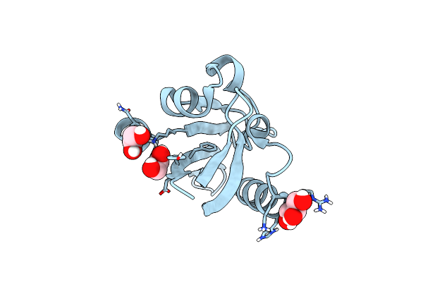
Organism: Homo sapiens
Method: X-RAY DIFFRACTION Resolution:1.31 Å Release Date: 2024-02-21 Classification: PROTEIN BINDING Ligands: GOL, PEG, CL, MG |
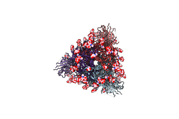 |
Organism: Severe acute respiratory syndrome coronavirus 2
Method: ELECTRON MICROSCOPY Release Date: 2023-06-07 Classification: VIRAL PROTEIN Ligands: NAG |
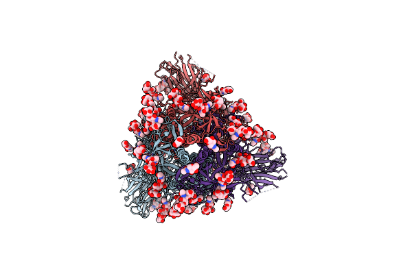 |
Organism: Severe acute respiratory syndrome coronavirus 2
Method: ELECTRON MICROSCOPY Release Date: 2023-06-07 Classification: VIRAL PROTEIN Ligands: NAG |
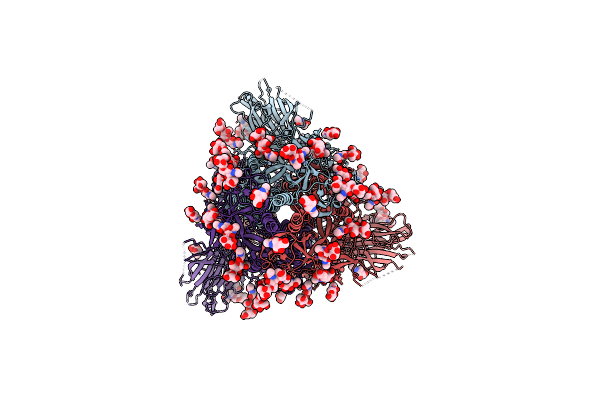 |
Organism: Severe acute respiratory syndrome coronavirus 2
Method: ELECTRON MICROSCOPY Release Date: 2023-06-07 Classification: VIRAL PROTEIN Ligands: NAG |
 |
Organism: Severe acute respiratory syndrome coronavirus 2, Mus musculus
Method: ELECTRON MICROSCOPY Release Date: 2023-04-05 Classification: VIRAL PROTEIN Ligands: NAG |
 |
Organism: Mus musculus, Severe acute respiratory syndrome coronavirus 2
Method: ELECTRON MICROSCOPY Release Date: 2023-04-05 Classification: VIRAL PROTEIN Ligands: NAG |
 |
Organism: Trypanosoma cruzi
Method: X-RAY DIFFRACTION Resolution:1.76 Å Release Date: 2022-06-22 Classification: OXIDOREDUCTASE Ligands: HEM, SO4, GOL, NA, OXY, ACT |
 |
Organism: Trypanosoma cruzi
Method: X-RAY DIFFRACTION Resolution:2.02 Å Release Date: 2022-06-08 Classification: OXIDOREDUCTASE Ligands: HEM, OXY, NA, SO4 |
 |
Organism: Severe acute respiratory syndrome coronavirus 2, Camelus dromedarius
Method: ELECTRON MICROSCOPY Release Date: 2022-04-20 Classification: VIRAL PROTEIN/IMMUNE SYSTEM |
 |
Crystal Structure Of Xanthomonas Campestris Tryptophan 2,3-Dioxygenase (Tdo)
Organism: Xanthomonas campestris pv. campestris (strain atcc 33913 / dsm 3586 / ncppb 528 / lmg 568 / p 25)
Method: X-RAY DIFFRACTION Resolution:1.70 Å Release Date: 2021-10-06 Classification: OXIDOREDUCTASE Ligands: HEM, CYN, KYN, TRP, GOL |
 |
Organism: Glycine max
Method: X-RAY DIFFRACTION Resolution:1.50 Å Release Date: 2021-04-21 Classification: OXIDOREDUCTASE Ligands: HEM, K |
 |
Organism: Saccharomyces cerevisiae
Method: X-RAY DIFFRACTION Resolution:1.06 Å Release Date: 2021-04-21 Classification: OXIDOREDUCTASE Ligands: HEC |
 |
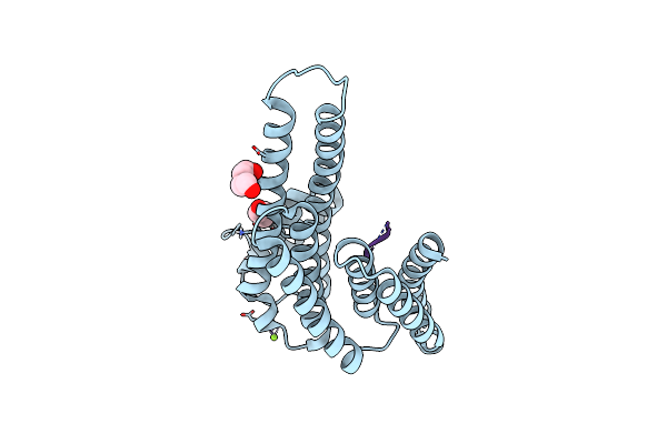
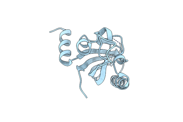
Crystal Structure Of The N-Terminal Pas Domain From The Herg3 Potassium Channel
Organism: Homo sapiens
Method: X-RAY DIFFRACTION Resolution:1.39 Å Release Date: 2020-08-12 Classification: MEMBRANE PROTEIN Ligands: GOL, EDO |
 |
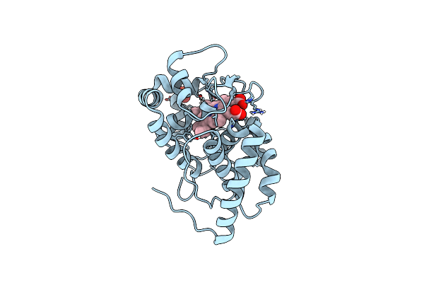
X-Ray Structure Of Human Glutamate Carboxypeptidase Ii (Gcpii) In Complex With A Inhibitor Kb1157
Organism: Homo sapiens
Method: X-RAY DIFFRACTION Resolution:1.77 Å Release Date: 2020-05-13 Classification: HYDROLASE Ligands: NAG, ZN, CA, CL, JX5 |
 |
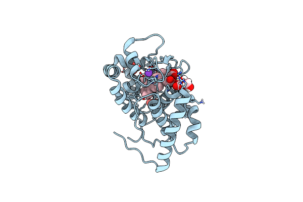
X-Ray Structure Of Human Glutamate Carboxypeptidase Ii (Gcpii)-E424M Inactive Mutant, In Complex With A Inhibitor Kb1160
Organism: Homo sapiens
Method: X-RAY DIFFRACTION Resolution:1.76 Å Release Date: 2020-05-13 Classification: HYDROLASE Ligands: NAG, ZN, CA, CL, KRZ |
 |
Organism: Glycine max
Method: X-RAY DIFFRACTION, NEUTRON DIFFRACTION Resolution:1.9000 Å, 2.2220 Å Release Date: 2020-03-18 Classification: OXIDOREDUCTASE Ligands: HEM, SO4, DOD |
 |
Organism: Glycine max
Method: X-RAY DIFFRACTION, NEUTRON DIFFRACTION Resolution:1.9000 Å, 2.0900 Å Release Date: 2020-03-18 Classification: OXIDOREDUCTASE Ligands: HEM, K, SO4, ASC, DOD |
 |
Organism: Homo sapiens
Method: X-RAY DIFFRACTION Resolution:2.32 Å Release Date: 2019-09-25 Classification: CIRCADIAN CLOCK PROTEIN |