Planned Maintenance: Some services may turn out to be unavailable from 15th January, 2026 to 16th January, 2026. We apologize for the inconvenience!

Planned Maintenance: Some services may turn out to be unavailable from 15th January, 2026 to 16th January, 2026. We apologize for the inconvenience!
 |
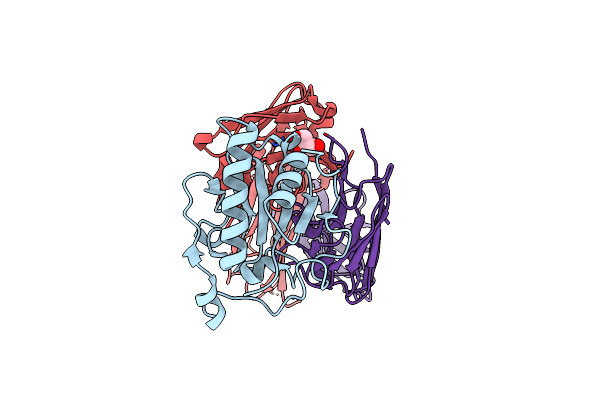
Organism: Vicugna pacos
Method: X-RAY DIFFRACTION Release Date: 2025-08-27 Classification: IMMUNE SYSTEM Ligands: SO4, CL, GOL, 1PE |
 |
Organism: Vicugna pacos
Method: X-RAY DIFFRACTION Release Date: 2025-08-27 Classification: IMMUNE SYSTEM Ligands: SO4, NA, GOL |
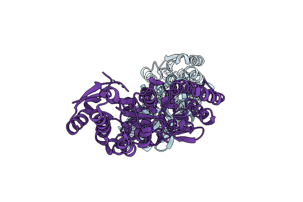 |
Crystal Structure Of Maltodextrin-Binding Protein Sps0871 From Streptococcus Pyogenes At 2.20 A
Organism: Streptococcus pyogenes m1 gas
Method: X-RAY DIFFRACTION Release Date: 2025-06-25 Classification: SUGAR BINDING PROTEIN |
 |
Crystal Structure Of Sonic Hedgehog In Complex With Antibody 5E1 Mutant H-R102A With Metals
Organism: Homo sapiens
Method: X-RAY DIFFRACTION Resolution:1.89 Å Release Date: 2025-04-16 Classification: IMMUNE SYSTEM Ligands: CL, ZN, CA, GOL |
 |
Crystal Structure Of Sonic Hedgehog In Complex With Antibody 5E1 Mutant L-T56A With Metals
Organism: Homo sapiens
Method: X-RAY DIFFRACTION Resolution:1.80 Å Release Date: 2025-04-16 Classification: IMMUNE SYSTEM Ligands: CA, ZN, CL, SO4 |
 |
Crystal Structure Of Sonic Hedgehog In Complex With Antibody 5E1 Without Metals
Organism: Homo sapiens
Method: X-RAY DIFFRACTION Resolution:1.75 Å Release Date: 2025-04-16 Classification: IMMUNE SYSTEM Ligands: GOL, NO3 |
 |
Crystal Structure Of Sonic Hedgehog In Complex With Antibody 5E1 Mutant H-R102A In The Absence Of Metals
Organism: Homo sapiens
Method: X-RAY DIFFRACTION Resolution:1.90 Å Release Date: 2025-04-09 Classification: IMMUNE SYSTEM Ligands: CL, GOL, IOD |
 |
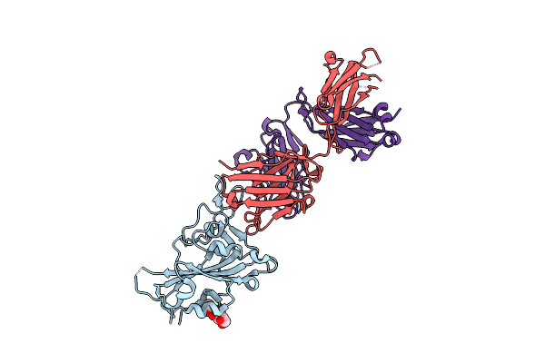
Crystal Structure Of The Anti-Phosphorylated Peptide C7 Scfv Antibody With Peptide Bound
Organism: Oryctolagus cuniculus, Homo sapiens
Method: X-RAY DIFFRACTION Resolution:2.80 Å Release Date: 2024-12-11 Classification: IMMUNE SYSTEM Ligands: SO4, CL |
 |
Crystal Structure Of The Anti-Phosphorylated Peptide C7 Fab Antibody With Peptide Bound
Organism: Oryctolagus cuniculus, Homo sapiens
Method: X-RAY DIFFRACTION Resolution:1.33 Å Release Date: 2024-12-11 Classification: IMMUNE SYSTEM Ligands: GOL, CL |
 |
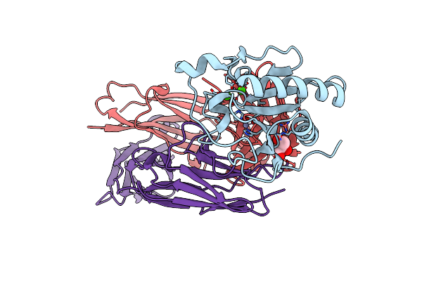
Organism: Severe acute respiratory syndrome coronavirus 2
Method: ELECTRON MICROSCOPY Release Date: 2024-10-09 Classification: VIRAL PROTEIN Ligands: NAG |
 |
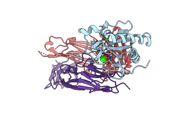
Organism: Severe acute respiratory syndrome coronavirus 2
Method: ELECTRON MICROSCOPY Release Date: 2024-10-09 Classification: VIRAL PROTEIN Ligands: NAG |
 |
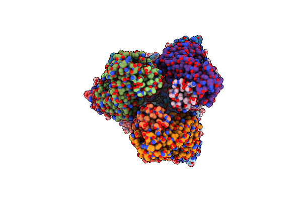
Structure Of Sars-Cov-2 Ba.2.86 Spike Glycoprotein In Complex With Ace2 (2-Up State)
Organism: Severe acute respiratory syndrome coronavirus 2, Homo sapiens
Method: ELECTRON MICROSCOPY Release Date: 2024-10-09 Classification: VIRAL PROTEIN/PROTEIN BINDING Ligands: NAG |
 |
Structure Of Sars-Cov-2 Ba.2.86 Spike Glycoprotein In Complex With Ace2 (2-Up And 1-Down State)
Organism: Severe acute respiratory syndrome coronavirus 2, Homo sapiens
Method: ELECTRON MICROSCOPY Release Date: 2024-10-09 Classification: VIRAL PROTEIN/PROTEIN BINDING Ligands: NAG |
 |
Organism: Homo sapiens, Severe acute respiratory syndrome coronavirus 2
Method: ELECTRON MICROSCOPY Release Date: 2024-10-09 Classification: VIRAL PROTEIN/PROTEIN BINDING Ligands: NAG |
 |
Structure Of Sars-Cov-2 Ba.2.86 Spike Rbd In Complex With Ace2 (Down State)
Organism: Severe acute respiratory syndrome coronavirus 2, Homo sapiens
Method: ELECTRON MICROSCOPY Release Date: 2024-10-09 Classification: VIRAL PROTEIN/PROTEIN BINDING Ligands: NAG |
 |
Structure Of Sars-Cov-2 Ba.2.86 Spike Glycoprotein In Complex With Ace2 (3-Up State)
Organism: Severe acute respiratory syndrome coronavirus 2, Homo sapiens
Method: ELECTRON MICROSCOPY Release Date: 2024-10-09 Classification: VIRAL PROTEIN/PROTEIN BINDING Ligands: NAG |
 |
Organism: Homo sapiens, Severe acute respiratory syndrome coronavirus 2
Method: ELECTRON MICROSCOPY Release Date: 2024-10-09 Classification: VIRAL PROTEIN/PROTEIN BINDING Ligands: NAG |
 |
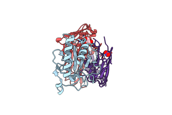
Organism: Oryctolagus cuniculus, Homo sapiens
Method: X-RAY DIFFRACTION Resolution:1.40 Å Release Date: 2024-06-12 Classification: IMMUNE SYSTEM Ligands: CL |
 |
Organism: Homo sapiens, Severe acute respiratory syndrome coronavirus 2
Method: X-RAY DIFFRACTION Resolution:2.20 Å Release Date: 2023-11-08 Classification: VIRAL PROTEIN/IMMUNE SYSTEM Ligands: NAG |
 |
Organism: Severe acute respiratory syndrome coronavirus 2, Homo sapiens
Method: ELECTRON MICROSCOPY Release Date: 2023-10-25 Classification: VIRAL PROTEIN/IMMUNE SYSTEM Ligands: NAG |