
Search Count: 30
 |
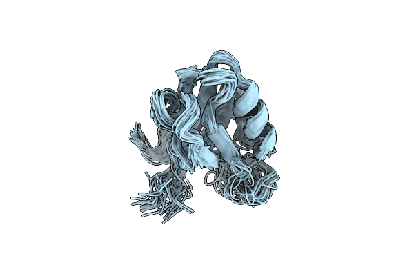
Organism: Hepatitis c virus (isolate 1)
Method: SOLUTION NMR Release Date: 2026-01-28 Classification: RNA |
 |
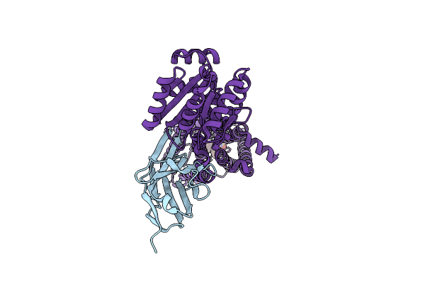
Organism: Plasmodium falciparum
Method: X-RAY DIFFRACTION Resolution:2.13 Å Release Date: 2025-05-21 Classification: STRUCTURAL PROTEIN |
 |
Structural Mechanism Of Cb1R Binding To Peripheral And Biased Inverse Agonists
Organism: Homo sapiens, Pyrococcus abyssi ge5, Lama glama
Method: ELECTRON MICROSCOPY Release Date: 2024-11-20 Classification: SIGNALING PROTEIN/IMMUNE SYSTEM Ligands: 7DY |
 |
Structural Mechanism Of Cb1R Binding To Peripheral And Biased Inverse Agonists
Organism: Lama glama, Homo sapiens, Pyrococcus abyssi ge5
Method: ELECTRON MICROSCOPY Release Date: 2024-11-20 Classification: SIGNALING PROTEIN/IMMUNE SYSTEM Ligands: A1AKO |
 |
Structural Mechanism Of Cb1R Binding To Peripheral And Biased Inverse Agonists
Organism: Homo sapiens, Pyrococcus abyssi ge5, Lama glama
Method: ELECTRON MICROSCOPY Release Date: 2024-11-20 Classification: SIGNALING PROTEIN/IMMUNE SYSTEM Ligands: A1AKN |
 |
Structure Of Duffy Antigen Receptor For Chemokines (Darc)/Ackr1 In Complex With The Chemokine, Ccl7 (Composite Map)
Organism: Homo sapiens
Method: ELECTRON MICROSCOPY Release Date: 2024-07-31 Classification: IMMUNE SYSTEM |
 |
Organism: Diploptera punctata
Method: X-RAY DIFFRACTION Resolution:2.95 Å Release Date: 2023-08-23 Classification: LIPID BINDING PROTEIN Ligands: NAG, ZN |
 |
Organism: Diploptera punctata
Method: X-RAY DIFFRACTION Resolution:2.10 Å Release Date: 2023-08-23 Classification: LIPID BINDING PROTEIN Ligands: NAG |
 |
Organism: Homo sapiens, Tequatrovirus t4
Method: ELECTRON MICROSCOPY Release Date: 2023-05-24 Classification: MEMBRANE PROTEIN Ligands: HXA |
 |
Organism: Homo sapiens
Method: ELECTRON MICROSCOPY Release Date: 2023-05-24 Classification: MEMBRANE PROTEIN Ligands: 2YB |
 |
Organism: Homo sapiens
Method: ELECTRON MICROSCOPY Release Date: 2023-05-24 Classification: MEMBRANE PROTEIN Ligands: 2YB |
 |
 |
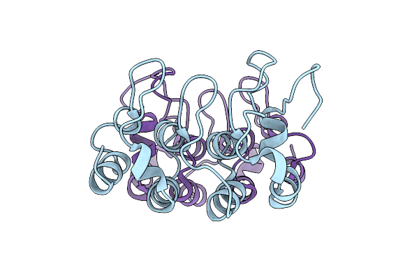
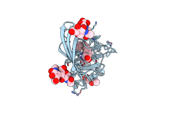
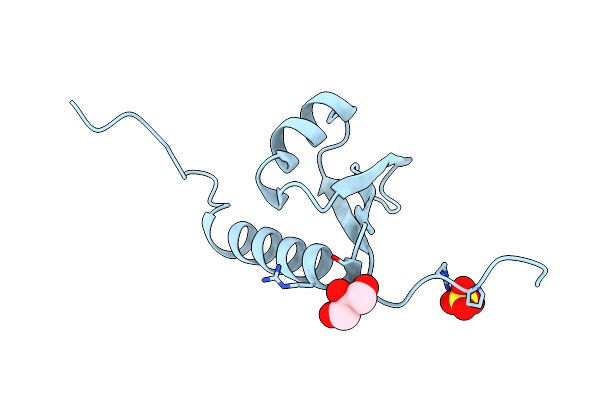
Two-State Liquid Nmr Structure Of A Pdz2 Domain From Hptp1E, Complexed With Ra-Gef2 Peptide
|
 |
Organism: Arabidopsis thaliana
Method: X-RAY DIFFRACTION Resolution:1.70 Å Release Date: 2022-04-13 Classification: PLANT PROTEIN Ligands: CA |
 |
Diploptera Punctata Inspired Lipocalin-Like Milk Protein Expressed In Saccharomyces Cerevisiae
Organism: Diploptera punctata
Method: X-RAY DIFFRACTION Resolution:2.35 Å Release Date: 2021-12-22 Classification: LIPID BINDING PROTEIN Ligands: NAG, PAM, GOL, ZN |
 |
Zn-Free Structure Of Lipocalin-Like Milk Protein, Inspired From Diploptera Punctata, Expressed In Saccharomyces Cerevisiae
Organism: Diploptera punctata
Method: X-RAY DIFFRACTION Resolution:1.45 Å Release Date: 2021-12-22 Classification: LIPID BINDING PROTEIN Ligands: PAM |
 |
Organism: Neurospora crassa or74a
Method: X-RAY DIFFRACTION Resolution:1.95 Å Release Date: 2021-06-30 Classification: DNA BINDING PROTEIN |
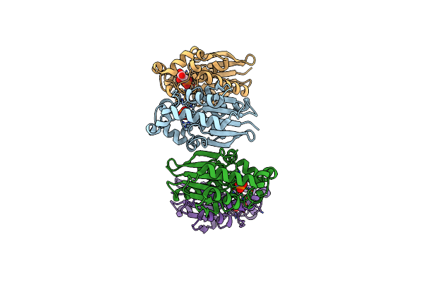 |
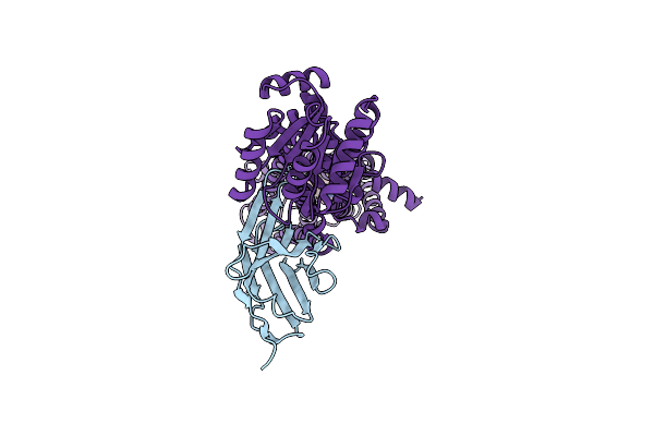
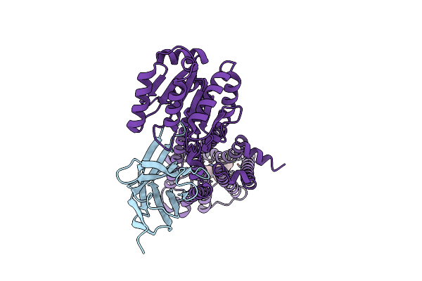
Crystal Structure Of Phosphoserine Phosphatase In Complex With 3-Phosphoglyceric Acid From Entamoeba Histolytica
Organism: Entamoeba histolytica
Method: X-RAY DIFFRACTION Resolution:2.48 Å Release Date: 2021-03-03 Classification: CYTOSOLIC PROTEIN Ligands: PO4, 3PG |
 |
Organism: Homo sapiens
Method: X-RAY DIFFRACTION Resolution:2.30 Å Release Date: 2020-12-02 Classification: TRANSCRIPTION Ligands: GOL, SO4 |
 |
Organism: Rattus norvegicus, Homo sapiens, Enterobacteria phage t4
Method: ELECTRON MICROSCOPY Release Date: 2020-06-17 Classification: SIGNALING PROTEIN Ligands: 08Y |