
Search Count: 15
 |
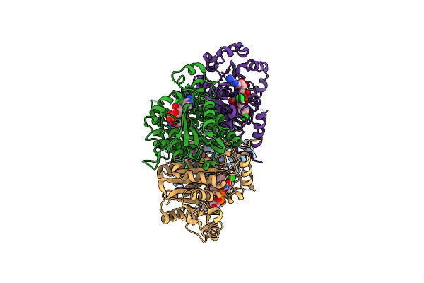
Organism: Sphaerobacter thermophilus (strain dsm 20745 / s 6022)
Method: X-RAY DIFFRACTION Resolution:2.10 Å Release Date: 2022-12-21 Classification: HYDROLASE |
 |
Crystal Structure Of Queuine Salvage Enzyme Duf2419 Complexed With Queuosine
Organism: Sphaerobacter thermophilus dsm 20745
Method: X-RAY DIFFRACTION Resolution:2.35 Å Release Date: 2022-12-21 Classification: HYDROLASE Ligands: QEO, MLI, PEG |
 |
Crystal Structure Of Queuine Salvage Enzyme Duf2419 Mutant K199C, Complexed With Queuosine
Organism: Sphaerobacter thermophilus dsm 20745
Method: X-RAY DIFFRACTION Resolution:2.50 Å Release Date: 2022-12-21 Classification: HYDROLASE Ligands: QEO, MLI |
 |
Crystal Structure Of Queuine Salvage Enzyme Duf2419, In Complex With Queuosine-5'-Monophosphate
Organism: Sphaerobacter thermophilus dsm 20745
Method: X-RAY DIFFRACTION Resolution:2.40 Å Release Date: 2022-12-21 Classification: HYDROLASE Ligands: 56B, NH4, PEG |
 |
Crystal Structure Of The Human Queuine Salvage Enzyme Duf2419, Wild-Type Apo Form
Organism: Homo sapiens
Method: X-RAY DIFFRACTION Resolution:1.78 Å Release Date: 2022-12-21 Classification: HYDROLASE Ligands: BTB |
 |
Crystal Structure Of Queuine Salvage Enzyme Duf2419, Wild-Type (Non-His6X Tagged)
Organism: Sphaerobacter thermophilus dsm 20745
Method: X-RAY DIFFRACTION Resolution:2.31 Å Release Date: 2022-12-21 Classification: HYDROLASE |
 |
Crystal Structure Of Queuine Salvage Enzyme Duf2419 Mutant D231N, In Complex With Queuosine-5'-Monophosphate
Organism: Sphaerobacter thermophilus dsm 20745
Method: X-RAY DIFFRACTION Resolution:1.99 Å Release Date: 2022-12-21 Classification: HYDROLASE Ligands: 56B, PEG |
 |
Crystal Structure Of The Human Queuine Salvage Enzyme Duf2419, Complexed With Queuine
Organism: Homo sapiens
Method: X-RAY DIFFRACTION Resolution:2.26 Å Release Date: 2022-12-21 Classification: HYDROLASE Ligands: QEI |
 |
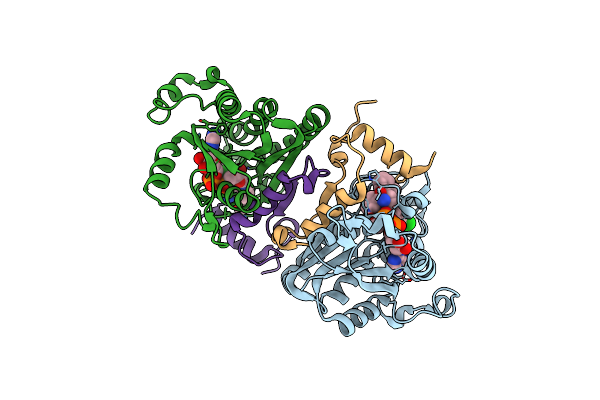
Crystal Structure Of Plasmodium Berghei Enoyl-Acyl-Carrier-Protein Reductase With Triclosan
Organism: Plasmodium berghei
Method: X-RAY DIFFRACTION Resolution:2.49 Å Release Date: 2010-03-02 Classification: OXIDOREDUCTASE Ligands: NAD, TCL |
 |
Organism: Plasmodium falciparum
Method: X-RAY DIFFRACTION Resolution:2.50 Å Release Date: 2007-07-17 Classification: OXIDOREDUCTASE Ligands: ZID |
 |
Crystal Structure Of Plasmodium Falciparum Enoyl Acp Reductase With Triclosan Reductase
Organism: Plasmodium falciparum
Method: X-RAY DIFFRACTION Resolution:2.26 Å Release Date: 2007-07-17 Classification: OXIDOREDUCTASE Ligands: NAD, JPN |
 |
Crystal Structure Of Plasmodium Falciparum Enoyl Acp Reductase With Triclosan Reductase
Organism: Plasmodium falciparum
Method: X-RAY DIFFRACTION Resolution:2.10 Å Release Date: 2007-07-17 Classification: OXIDOREDUCTASE Ligands: NAD, JPJ |
 |
Crystal Structure Of Plasmodium Falciparum Enoyl Acp Reductase With Triclosan Reductase
Organism: Plasmodium falciparum
Method: X-RAY DIFFRACTION Resolution:2.80 Å Release Date: 2007-07-17 Classification: OXIDOREDUCTASE Ligands: SO4, NAD, 7PC |
 |
Crystal Structure Of Plasmodium Falciparum Enoyl Acp Reductase With Triclosan Reductase
Organism: Plasmodium falciparum
Method: X-RAY DIFFRACTION Resolution:2.60 Å Release Date: 2007-07-17 Classification: OXIDOREDUCTASE Ligands: NAD, 8PC |
 |
Synthesis, Biological Activity, And X-Ray Crystal Structural Analysis Of Diaryl Ether Inhibitors Of Malarial Enoyl Acp Reductase.
Organism: Plasmodium falciparum
Method: X-RAY DIFFRACTION Resolution:2.50 Å Release Date: 2007-01-16 Classification: OXIDOREDUCTASE Ligands: NAD, JPA |