
Search Count: 271
 |
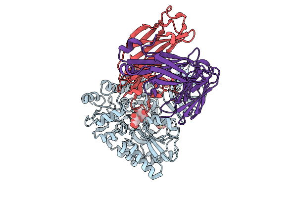
Crystal Structure Of Gossypium Hirsutum (Cotton) 5-Enol-Pyruvyl-Shikimate-3-Phosphate Synthase (Epsps) In Open Conformation
Organism: Gossypium hirsutum
Method: X-RAY DIFFRACTION Resolution:2.20 Å Release Date: 2026-01-07 Classification: TRANSFERASE Ligands: PEG, EDO, GOL, GPJ |
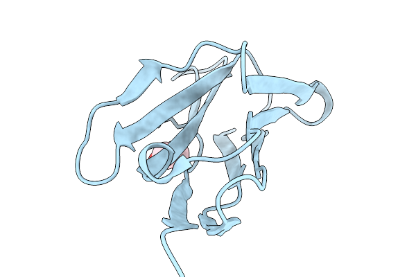 |
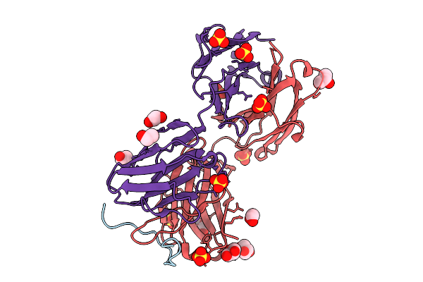
Organism: Chiloscyllium plagiosum
Method: X-RAY DIFFRACTION Resolution:2.00 Å Release Date: 2026-01-07 Classification: IMMUNE SYSTEM Ligands: EDO |
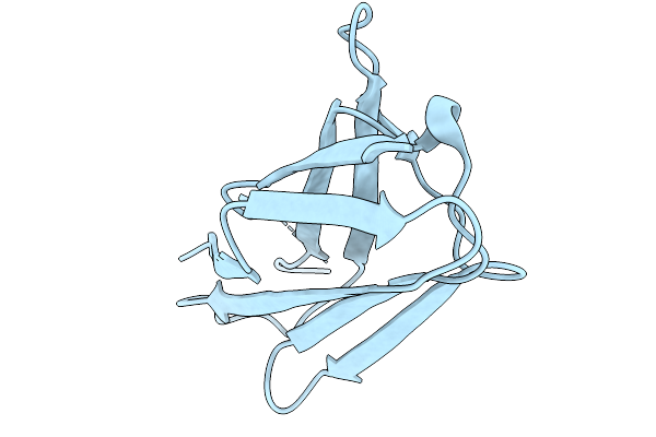 |
Organism: Chiloscyllium plagiosum
Method: X-RAY DIFFRACTION Resolution:1.80 Å Release Date: 2026-01-07 Classification: IMMUNE SYSTEM |
 |
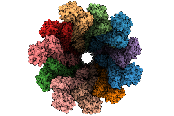
Organism: Homo sapiens
Method: ELECTRON MICROSCOPY Release Date: 2025-10-29 Classification: NEUROPEPTIDE |
 |
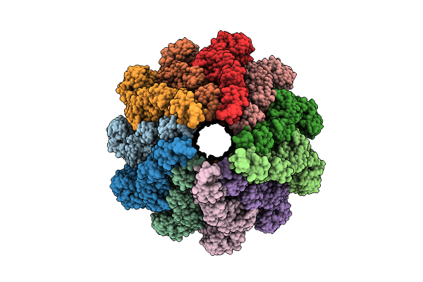
Organism: Homo sapiens
Method: ELECTRON MICROSCOPY Release Date: 2025-10-29 Classification: NEUROPEPTIDE |
 |
In Situ Structure Of The Sheathed Flad Flagellar Filament In Vibrio Cholerae
Organism: Vibrio cholerae o1 biovar el tor str. n16961
Method: ELECTRON MICROSCOPY Release Date: 2025-09-24 Classification: MOTOR PROTEIN |
 |
In Situ Unsheathed Flagellar Filament Of Vibrio Cholerae Resolved With Helical Reconstruction.
Organism: Vibrio cholerae o1 biovar el tor str. n16961
Method: ELECTRON MICROSCOPY Release Date: 2025-09-24 Classification: MOTOR PROTEIN |
 |
Organism: Vibrio cholerae o1 biovar el tor str. n16961
Method: ELECTRON MICROSCOPY Release Date: 2025-09-24 Classification: MOTOR PROTEIN |
 |
In Situ Sheathed Flagellar Filament Of Vibrio Cholerae Resolved With Helical Reconstruction.
Organism: Vibrio cholerae o1 biovar el tor str. n16961
Method: ELECTRON MICROSCOPY Release Date: 2025-09-24 Classification: MOTOR PROTEIN |
 |
Organism: Vibrio cholerae o1 biovar el tor str. n16961
Method: ELECTRON MICROSCOPY Release Date: 2025-09-24 Classification: MOTOR PROTEIN |
 |
In Situ Structure Of The Sheathed Flab Flagellar Filament In Vibrio Cholerae
Organism: Vibrio cholerae o1 biovar el tor
Method: ELECTRON MICROSCOPY Release Date: 2025-09-24 Classification: MOTOR PROTEIN |
 |
Organism: Homo sapiens
Method: X-RAY DIFFRACTION Resolution:1.80 Å Release Date: 2025-09-10 Classification: IMMUNE SYSTEM Ligands: PEG, GOL, SO4, EDO |
 |
Organism: Escherichia coli, Homo sapiens
Method: X-RAY DIFFRACTION Resolution:3.70 Å Release Date: 2025-09-10 Classification: IMMUNE SYSTEM Ligands: SO4 |
 |
Organism: Homo sapiens
Method: X-RAY DIFFRACTION Resolution:1.14 Å Release Date: 2025-04-30 Classification: METAL BINDING PROTEIN Ligands: ZN, CO2 |
 |
Organism: Homo sapiens
Method: X-RAY DIFFRACTION Resolution:1.20 Å Release Date: 2025-04-30 Classification: METAL BINDING PROTEIN Ligands: ZN, MNP |
 |
Organism: Homo sapiens
Method: X-RAY DIFFRACTION Resolution:1.20 Å Release Date: 2025-04-30 Classification: METAL BINDING PROTEIN Ligands: ZN, MNP |
 |
Organism: Homo sapiens
Method: X-RAY DIFFRACTION Resolution:1.20 Å Release Date: 2025-04-30 Classification: METAL BINDING PROTEIN Ligands: ZN, MNP |
 |
Organism: Homo sapiens
Method: X-RAY DIFFRACTION Resolution:1.20 Å Release Date: 2025-04-30 Classification: METAL BINDING PROTEIN Ligands: ZN, MNP |
 |
Organism: Homo sapiens
Method: X-RAY DIFFRACTION Resolution:1.20 Å Release Date: 2025-04-30 Classification: METAL BINDING PROTEIN Ligands: ZN, MNP |
 |
Organism: Homo sapiens
Method: X-RAY DIFFRACTION Resolution:1.35 Å Release Date: 2025-04-30 Classification: METAL BINDING PROTEIN Ligands: ZN, MNP |