
Search Count: 27
 |
Organism: Mangifera indica
Method: X-RAY DIFFRACTION Resolution:2.40 Å Release Date: 2024-08-07 Classification: PLANT PROTEIN Ligands: PG4 |
 |
Organism: Santalum album
Method: X-RAY DIFFRACTION Resolution:3.41 Å Release Date: 2022-03-09 Classification: LYASE Ligands: MG |
 |
Organism: Santalum album
Method: X-RAY DIFFRACTION Resolution:3.10 Å Release Date: 2022-03-02 Classification: LYASE |
 |
Crystal Structure Of Porcine Pancreatic Trypsin With Tripeptide Inhibitor, Prn, At Ph10
Organism: Capsicum annuum, Sus scrofa
Method: X-RAY DIFFRACTION Resolution:1.90 Å Release Date: 2018-07-04 Classification: HYDROLASE/HYDROLASE INHIBITOR Ligands: CA, MPD |
 |
Crystal Structure Of Porcine Pancreatic Trypsin With Tripeptide Inhibitor, Prn, At Ph 7
Organism: Capsicum annuum, Sus scrofa
Method: X-RAY DIFFRACTION Resolution:2.00 Å Release Date: 2018-03-28 Classification: HYDROLASE/HYDROLASE INHIBITOR Ligands: CA, MPD |
 |
Crystal Structure Of Porcine Pancreatic Trypsin With Tripeptide Inhibitor, Pry, At Ph 7
Organism: Capsicum annuum, Sus scrofa
Method: X-RAY DIFFRACTION Resolution:2.00 Å Release Date: 2018-03-28 Classification: HYDROLASE/HYDROLASE INHIBITOR Ligands: CA |
 |
Crystal Structure Of Porcine Pancreatic Trypsin With Tripeptide Inhibitor, Pry, At Ph 10
Organism: Capsicum annuum, Sus scrofa
Method: X-RAY DIFFRACTION Resolution:1.90 Å Release Date: 2018-03-28 Classification: HYDROLASE/HYDROLASE INHIBITOR Ligands: CA |
 |
Crystal Structure Of Porcine Pancreatic Trypsin With Tripeptide Inhibitor, Tre, At Ph 7
Organism: Capsicum annuum, Sus scrofa
Method: X-RAY DIFFRACTION Resolution:1.80 Å Release Date: 2018-03-28 Classification: HYDROLASE/HYDROLASE INHIBITOR Ligands: CA, MPD |
 |
Crystal Structure Of Porcine Pancreatic Trypsin With Tripeptide Inhibitor, Tre, At Ph 10
Organism: Capsicum annuum, Sus scrofa
Method: X-RAY DIFFRACTION Resolution:2.10 Å Release Date: 2018-03-28 Classification: HYDROLASE/HYDROLASE INHIBITOR Ligands: CA, MPD |
 |
Organism: Schizosaccharomyces pombe
Method: X-RAY DIFFRACTION Resolution:2.30 Å Release Date: 2012-03-21 Classification: CELL CYCLE |
 |
Organism: Schizosaccharomyces pombe
Method: X-RAY DIFFRACTION Resolution:2.60 Å Release Date: 2011-03-30 Classification: CELL CYCLE Ligands: AUC |
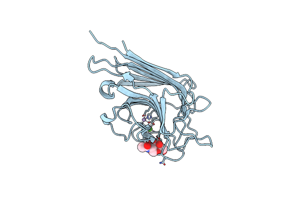 |
Organism: Erythrina corallodendron
Method: X-RAY DIFFRACTION Resolution:2.00 Å Release Date: 2011-03-30 Classification: SUGAR BINDING PROTEIN Ligands: MN, CA, A2G |
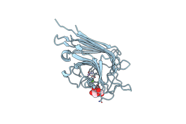 |
Organism: Erythrina corallodendron
Method: X-RAY DIFFRACTION Resolution:2.30 Å Release Date: 2011-03-30 Classification: SUGAR BINDING PROTEIN Ligands: MN, CA, GLA |
 |
Organism: Erythrina corallodendron
Method: X-RAY DIFFRACTION Resolution:2.00 Å Release Date: 2011-03-30 Classification: SUGAR BINDING PROTEIN Ligands: MN, CA, CIT |
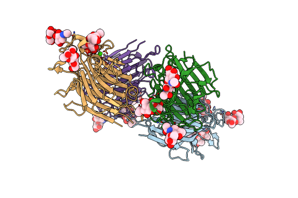 |
Crystl Structure Of Basic Winged Bean Lectin In Complex With Gal-Alpha-1,4-Gal-Beta-Ethylene
Organism: Psophocarpus tetragonolobus
Method: X-RAY DIFFRACTION Resolution:2.50 Å Release Date: 2008-07-29 Classification: SUGAR BINDING PROTEIN Ligands: MN, CA, NAG, BMA |
 |
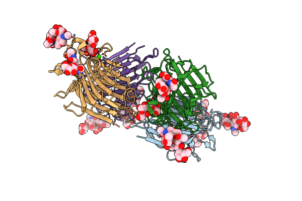
Crystal Structure Of Basic Winged Bean Lectin In Complex With Gal-Alpha 1,4 Gal
Organism: Psophocarpus tetragonolobus
Method: X-RAY DIFFRACTION Resolution:2.65 Å Release Date: 2008-07-29 Classification: SUGAR BINDING PROTEIN Ligands: NAG, MN, CA |
 |
Crystal Structure Of Basic Winged Bean Lectin In Complex With Gal-Alpha- 1,6 Glc
Organism: Psophocarpus tetragonolobus
Method: X-RAY DIFFRACTION Resolution:2.90 Å Release Date: 2008-07-29 Classification: SUGAR BINDING PROTEIN Ligands: MN, CA |
 |
Crystal Structure Of Basic Winged Bean Lectin In Complex With A Blood Group Disaccharide
Organism: Psophocarpus tetragonolobus
Method: X-RAY DIFFRACTION Resolution:2.50 Å Release Date: 2007-06-26 Classification: SUGAR BINDING PROTEIN Ligands: MN, CA |
 |
Crystal Structure Of Basic Winged Bean Lectin In Complex With B Blood Group Disaccharide
Organism: Psophocarpus tetragonolobus
Method: X-RAY DIFFRACTION Resolution:2.40 Å Release Date: 2007-06-26 Classification: SUGAR BINDING PROTEIN Ligands: CA, MN |
 |
Crystal Structure Of Basic Winged Bean Lectin In Complex With B Blood Group Trisaccharide
Organism: Psophocarpus tetragonolobus
Method: X-RAY DIFFRACTION Resolution:2.75 Å Release Date: 2007-06-19 Classification: SUGAR BINDING PROTEIN Ligands: CA, MN |