
Search Count: 15
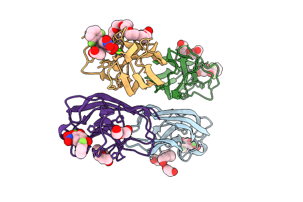 |
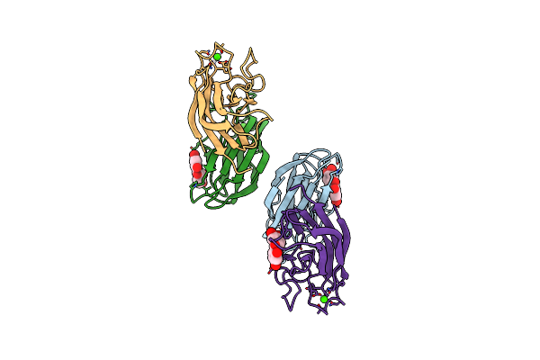
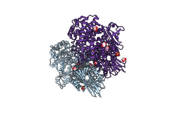
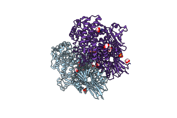
Organism: Pseudomonas aeruginosa pao1
Method: X-RAY DIFFRACTION Release Date: 2025-10-22 Classification: SUGAR BINDING PROTEIN Ligands: CA, A1IZM, 1PE, EDO, CL, PG4, 9YU, ACT, PEG |
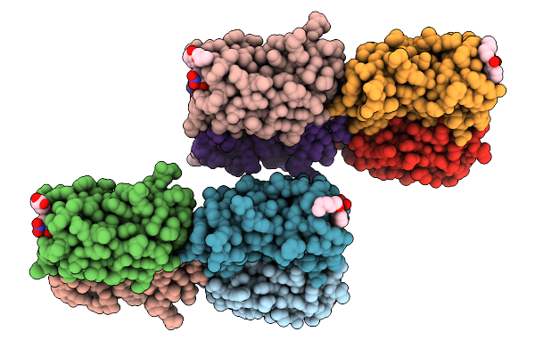 |
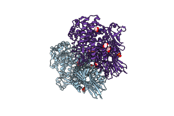
Organism: Pseudomonas aeruginosa pao1
Method: X-RAY DIFFRACTION Release Date: 2025-10-22 Classification: SUGAR BINDING PROTEIN Ligands: CA, A1I1G |
 |
Leca From Pseudomonas Aeruginosa In Complex With Tolcapone (Cas: 134308-13-7)
Organism: Pseudomonas aeruginosa
Method: X-RAY DIFFRACTION Resolution:1.32 Å Release Date: 2023-07-19 Classification: SUGAR BINDING PROTEIN Ligands: CA, TCW |
 |
Leca From Pseudomonas Aeruginosa In Complex With 4-Phenylbutyryl Hydroxamic Acid (Cas: 32153-46-1)
Organism: Pseudomonas aeruginosa
Method: X-RAY DIFFRACTION Resolution:1.79 Å Release Date: 2022-06-08 Classification: SUGAR BINDING PROTEIN Ligands: 4R9, CA |
 |
Leca From Pseudomonas Aeruginosa In Complex With A Synthetic Monovalent Galactosidic Inhibitor
Organism: Pseudomonas aeruginosa
Method: X-RAY DIFFRACTION Resolution:1.50 Å Release Date: 2022-02-16 Classification: SUGAR BINDING PROTEIN Ligands: CA, 4VH, EDO |
 |
Leca From Pseudomonas Aeruginosa In Complex With A Catechol Cas No. 61445-50-9
Organism: Pseudomonas aeruginosa pao1
Method: X-RAY DIFFRACTION Resolution:1.84 Å Release Date: 2021-02-24 Classification: SUGAR BINDING PROTEIN Ligands: CA, P4H |
 |
Leca From Pseudomonas Aeruginosa In Complex With A Catechol Cas No. 67984-81-0
Organism: Pseudomonas aeruginosa (strain atcc 15692 / dsm 22644 / cip 104116 / jcm 14847 / lmg 12228 / 1c / prs 101 / pao1)
Method: X-RAY DIFFRACTION Resolution:1.84 Å Release Date: 2020-12-30 Classification: SUGAR BINDING PROTEIN Ligands: CA, P3K, P6G, EDO, PG4, PGE |
 |
Fucose-Binding Lectin From Burkholderia Ambifaria (Bambl) In Complex With A Fucosyl Derivative
Organism: Burkholderia ambifaria (strain atcc baa-244 / ammd)
Method: X-RAY DIFFRACTION Resolution:1.65 Å Release Date: 2020-10-28 Classification: SUGAR BINDING PROTEIN Ligands: QJB |
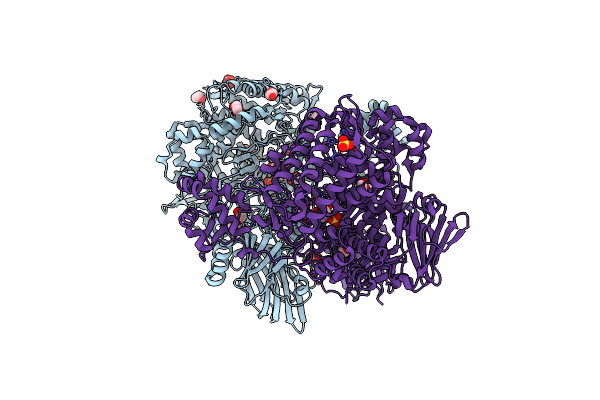 |
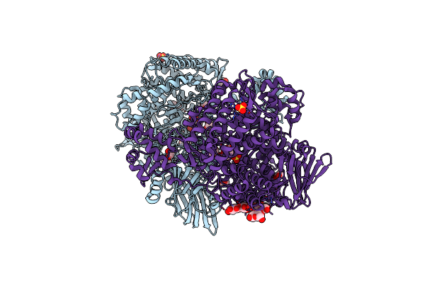
Organism: Metagenome
Method: X-RAY DIFFRACTION Resolution:2.05 Å Release Date: 2019-06-12 Classification: HYDROLASE Ligands: SO4, BCN, EDO, CL |
 |
Bacterial Beta-1,3-Oligosaccharide Phosphorylase From Gh149 With Laminarihexaose Bound At A Surface Site
Organism: Metagenome
Method: X-RAY DIFFRACTION Resolution:2.25 Å Release Date: 2019-06-12 Classification: HYDROLASE Ligands: SO4, BCN, EDO, CL |
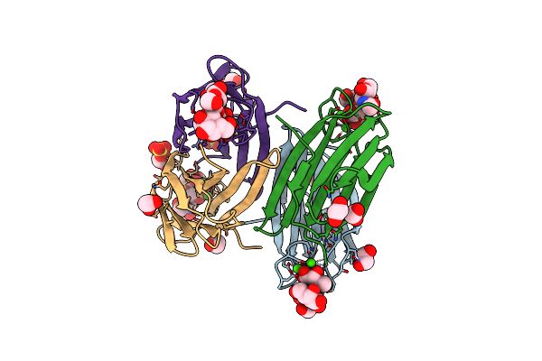 |
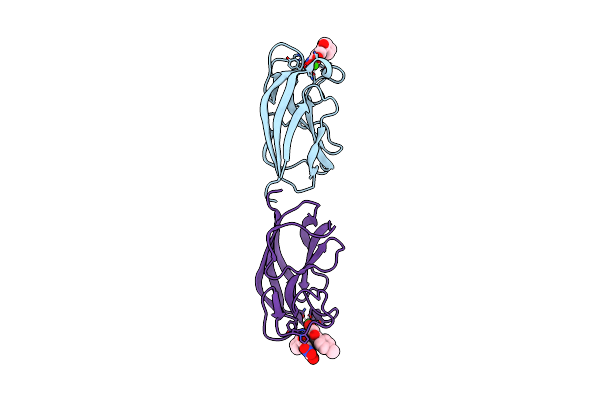
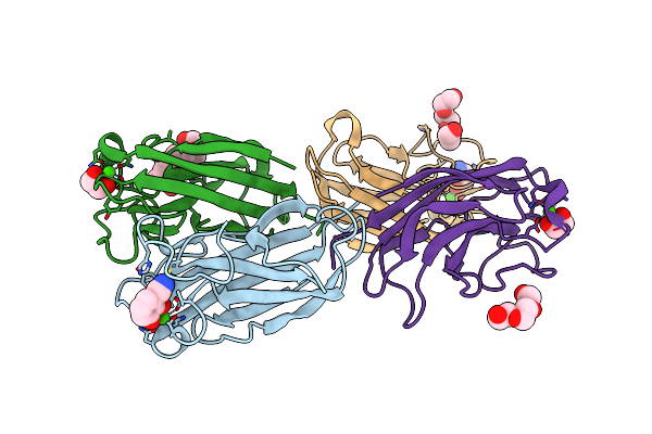
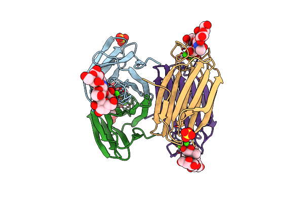
Structure Of The Lecb Lectin From Pseudomonas Aeruginosa Strain Pao1 In Complex With Lewis X Tetrasaccharide
Organism: Pseudomonas aeruginosa pao1
Method: X-RAY DIFFRACTION Resolution:1.80 Å Release Date: 2019-06-12 Classification: SUGAR BINDING PROTEIN Ligands: CA, SO4, EDO |
 |
Organism: Paenibacillus sp. ym1
Method: X-RAY DIFFRACTION Resolution:1.95 Å Release Date: 2018-06-13 Classification: HYDROLASE Ligands: SO4, EDO, CL |
 |
Paenibacillus Sp. Ym1 Laminaribiose Phosphorylase With Alpha-Glc-1-Phosphate Bound
Organism: Paenibacillus sp. ym1
Method: X-RAY DIFFRACTION Resolution:2.50 Å Release Date: 2018-06-13 Classification: HYDROLASE Ligands: SO4, G1P, EDO, CL |
 |
Paenibacillus Sp. Ym1 Laminaribiose Phosphorylase With Alpha-Man-1-Phosphate Bound
Organism: Paenibacillus sp. ym1
Method: X-RAY DIFFRACTION Resolution:1.82 Å Release Date: 2018-06-13 Classification: HYDROLASE Ligands: SO4, M1P, EDO, CL |
 |
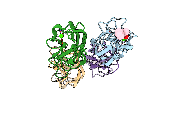
Structure Of The Lecb Lectin From Pseudomonas Aeruginosa Strain Pa14 In Complex With Lewis X Tetrasaccharide
Organism: Pseudomonas aeruginosa
Method: X-RAY DIFFRACTION Resolution:1.60 Å Release Date: 2016-07-27 Classification: SUGAR BINDING PROTEIN Ligands: CA, SO4 |