
Search Count: 49
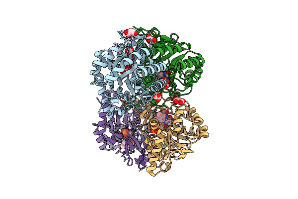 |
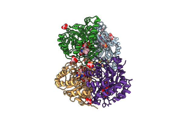
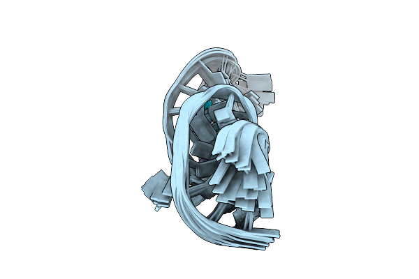
Crystal Structure Of The C131A Mutant Of Trypanosoma Brucei Dhodh In Fmn-Oxidized, Ligand-Free Form
Organism: Trypanosoma brucei brucei treu927
Method: X-RAY DIFFRACTION Release Date: 2025-11-26 Classification: FLAVOPROTEIN Ligands: FMN, MLI |
 |
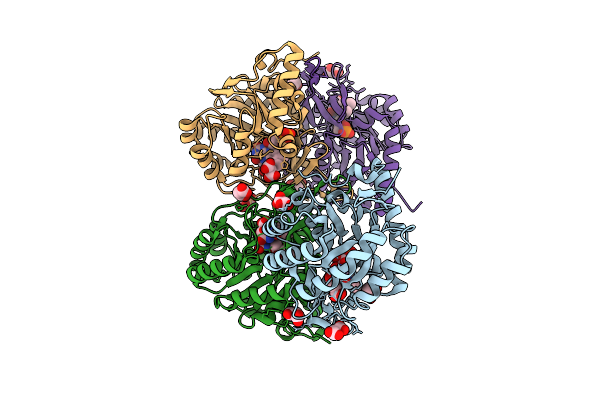
Crystal Structure Of The C131A Mutant Of Trypanosoma Brucei Dhodh In Fmn-Reduced, Ligand-Free Form
Organism: Trypanosoma brucei brucei treu927
Method: X-RAY DIFFRACTION Release Date: 2025-11-26 Classification: FLAVOPROTEIN Ligands: FNR, MLI, SO3 |
 |
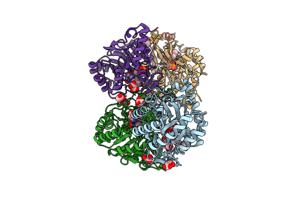
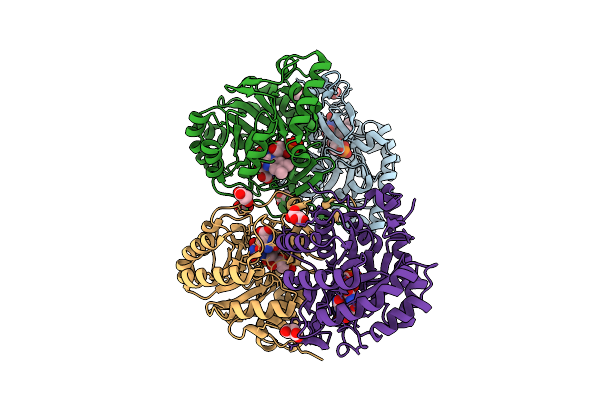
Crystal Structure Of The C131A Mutant Of Trypanosoma Brucei Dhodh In Fmn-Oxidized, Dihydroorotate-Bound Form
Organism: Trypanosoma brucei brucei treu927
Method: X-RAY DIFFRACTION Release Date: 2025-11-26 Classification: FLAVOPROTEIN Ligands: FMN, DOR, MLI |
 |
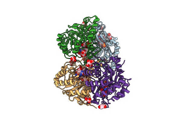
Crystal Structure Of The C131A Mutant Of Trypanosoma Brucei Dhodh In Fmn-Reduced, Dihydroorotate-Bound Form
Organism: Trypanosoma brucei brucei treu927
Method: X-RAY DIFFRACTION Release Date: 2025-11-26 Classification: FLAVOPROTEIN Ligands: FNR, DOR, MLI |
 |
Crystal Structure Of The C131A Mutant Of Trypanosoma Brucei Dhodh In Fmn-Oxidized, Orotate-Bound Form
Organism: Trypanosoma brucei brucei treu927
Method: X-RAY DIFFRACTION Release Date: 2025-11-26 Classification: FLAVOPROTEIN Ligands: FMN, ORO, MLI |
 |
Crystal Structure Of The C131A Mutant Of Trypanosoma Brucei Dhodh In Fmn-Reduced, Orotate-Bound Form
Organism: Trypanosoma brucei brucei treu927
Method: X-RAY DIFFRACTION Release Date: 2025-11-26 Classification: FLAVOPROTEIN Ligands: FNR, ORO, MLI |
 |
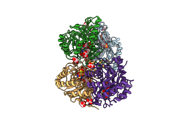
Crystal Structure Of The C131A Mutant Of Trypanosoma Brucei Dhodh In Fmn-Oxidized, Fumarate-Bound Form
Organism: Trypanosoma brucei brucei treu927
Method: X-RAY DIFFRACTION Release Date: 2025-11-26 Classification: FLAVOPROTEIN Ligands: FMN, FUM, MLI |
 |
Crystal Structure Of The C131A Mutant Of Trypanosoma Brucei Dhodh In Fmn-Reduced, Fumarate-Bound Form
Organism: Trypanosoma brucei brucei treu927
Method: X-RAY DIFFRACTION Release Date: 2025-11-26 Classification: FLAVOPROTEIN Ligands: FNR, FUM, MLI |
 |
Crystal Structure Of Wild-Type Trypanosoma Brucei Dhodh In Fmn-Reduced, Ligand-Free Form
Organism: Trypanosoma brucei brucei treu927
Method: X-RAY DIFFRACTION Release Date: 2025-11-26 Classification: FLAVOPROTEIN Ligands: FNR, MLI, SO2, SO3 |
 |
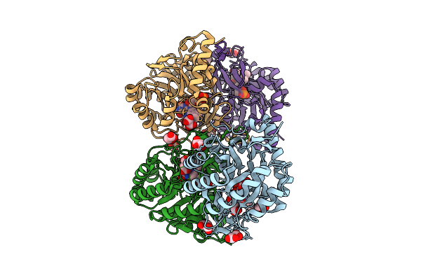
Crystal Structure Of Wild-Type Trypanosoma Brucei Dhodh In Fmn-Oxidized, Orotate-Bound Form
Organism: Trypanosoma brucei brucei treu927
Method: X-RAY DIFFRACTION Release Date: 2025-11-26 Classification: FLAVOPROTEIN Ligands: FMN, ORO, MLI |
 |
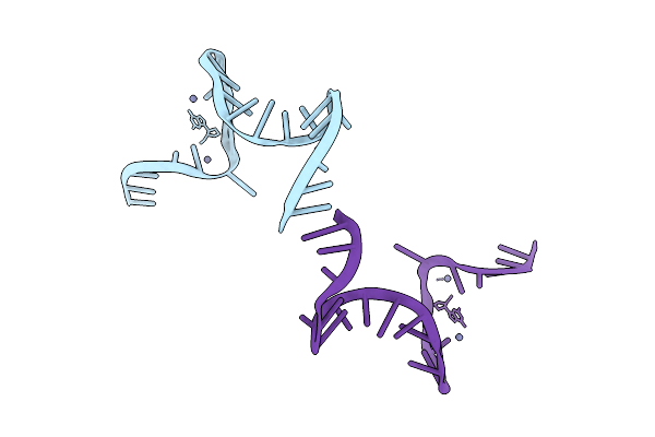
Organism: Synthetic construct
Method: SOLUTION NMR Release Date: 2025-10-29 Classification: DNA-RNA HYBRID Ligands: ZN |
 |
Organism: Synthetic construct
Method: X-RAY DIFFRACTION Release Date: 2025-10-29 Classification: DNA-RNA HYBRID Ligands: ZN |
 |
Organism: Synthetic construct
Method: X-RAY DIFFRACTION Release Date: 2025-10-29 Classification: DNA-RNA HYBRID Ligands: ZN |
 |
Organism: Oryza sativa
Method: X-RAY DIFFRACTION Release Date: 2025-06-25 Classification: OXIDOREDUCATASE/INHIBITOR Ligands: CO, A1L59, IOD |
 |
Crystal Structure Of Arabidopsis Thaliana Hppd Complexed With Iptriazopyrid
Organism: Arabidopsis thaliana
Method: X-RAY DIFFRACTION Release Date: 2025-06-25 Classification: OXIDOREDUCTASE Ligands: CO, A1L59 |
 |
Organism: Oryza sativa
Method: X-RAY DIFFRACTION Release Date: 2025-06-25 Classification: OXIDOREDUCTASE Ligands: CO, ACY, ZN |
 |
Organism: Burkholderia stagnalis
Method: X-RAY DIFFRACTION Resolution:1.40 Å Release Date: 2025-01-22 Classification: HYDROLASE Ligands: CA, SO4, MPD |
 |
Pholiota Squarrosa Lectin (Phosl) In Complex With Fucose(Alpha1-6)[Glcnac(Beta1-4)]Glcnac
Organism: Pholiota squarrosa
Method: X-RAY DIFFRACTION Resolution:2.15 Å Release Date: 2022-10-12 Classification: SUGAR BINDING PROTEIN Ligands: FMT |
 |
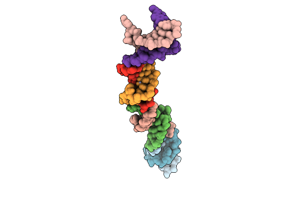
The Crystal Structure Of The Immature Apo-Enzyme Of Homoserine Dehydrogenase From The Hyperthermophilic Archaeon Sulfurisphaera Tokodaii.
Organism: Sulfurisphaera tokodaii
Method: X-RAY DIFFRACTION Resolution:2.05 Å Release Date: 2022-06-22 Classification: OXIDOREDUCTASE Ligands: MG |
 |
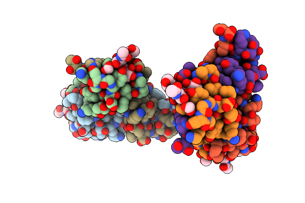
The Crystal Structure Of The Immature Holo-Enzyme Of Homoserine Dehydrogenase Complexed With Nadp And 1,4-Butandiol From The Hyperthermophilic Archaeon Sulfurisphaera Tokodaii.
Organism: Sulfurisphaera tokodaii (strain dsm 16993 / jcm 10545 / nbrc 100140 / 7)
Method: X-RAY DIFFRACTION Resolution:1.90 Å Release Date: 2022-06-22 Classification: OXIDOREDUCTASE Ligands: NAP, BU1 |